
-
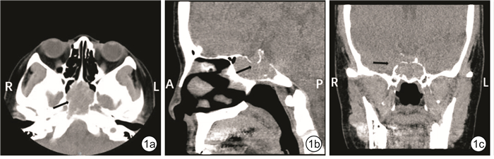
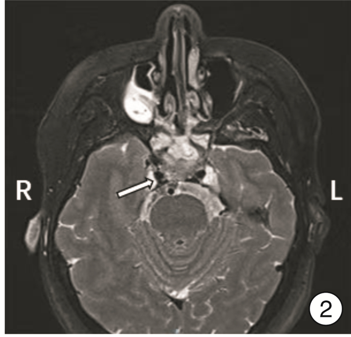
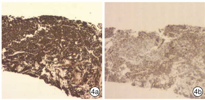
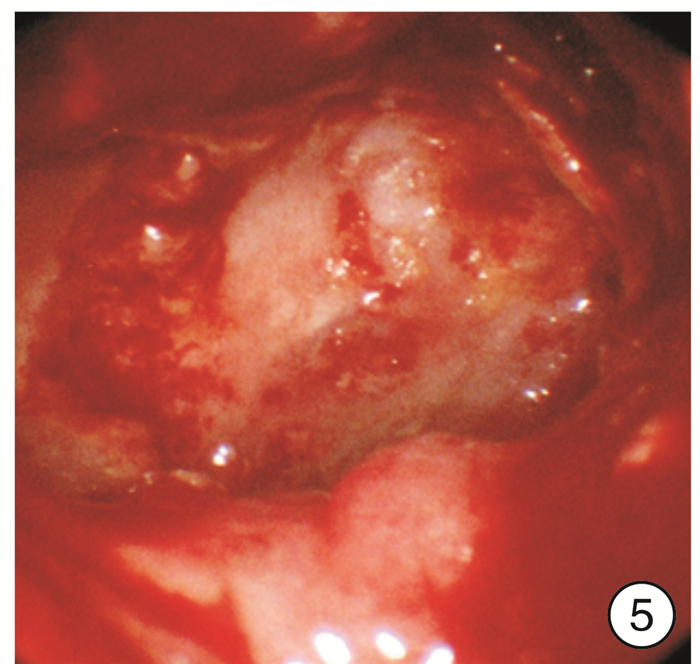
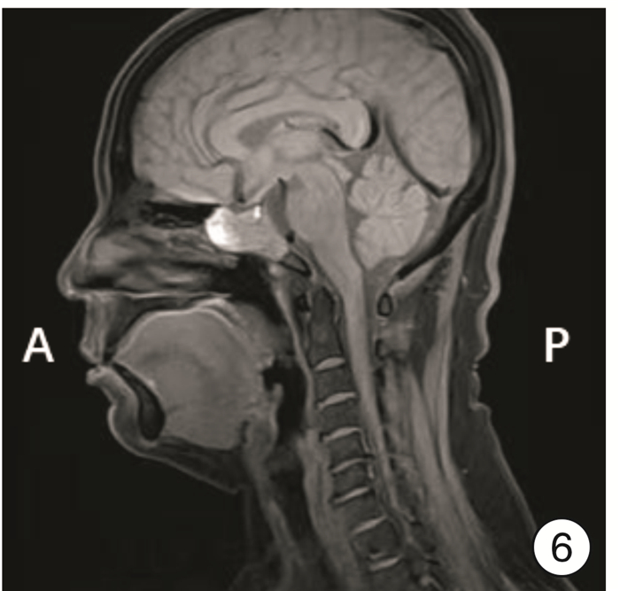
摘要: 异位垂体腺瘤在临床上较为罕见,本文报告1例蝶窦异位垂体腺瘤病例,总结其病例特点,并梳理其诊疗过程。54岁女性患者,因“体检发现鼻窦肿物1个月余,偶伴头胀,无头晕及头痛”就诊,影像学检查提示枕骨斜坡、双侧蝶窦内占位。患者于全身麻醉下行鼻内镜下经蝶窦脑病损切除术。术后病理提示为垂体神经内分泌肿瘤。术后恢复良好,无并发症,随访2个月,肿瘤未见复发。Abstract: Ectopic pituitary adenoma is rare in clinical practice. This article reports a case of ectopic pituitary adenoma of sphenoid sinus, and summarizes the clinical characteristics, diagnosis and management. A 54-year-old female patient complaining with occasional head distension without dizziness and headache for more than 1 month was admitted due to sinus mass on conventional physical examination. Imaging examination revealed a mass in the occipital slope and bilateral sphenoid sinus. The patient underwent endoscopic resection of the mass under general anesthesia. Postoperative histopathological examination showed "pituitary neuroendocrine tumor". Postoperative recovery was good and no complications occurred. She was followed up for 2 months without relapse.
-
Key words:
- sphenoid sinus /
- ectopic pituitary adenoma /
- pituitary tumor
-

-
[1] 孙永青, 许洋, 张林医, 等. 蝶窦异位垂体瘤二例报告并文献复习[J]. 解放军医药杂志, 2011, 23(S1): 34-34. https://www.cnki.com.cn/Article/CJFDTOTAL-HBGF2011S1039.htm
[2] 王艳, 姚新宇, 张德江, 等. 1例蝶窦异位垂体瘤影像学表现[J]. 中国介入影像与治疗学, 2023, 20(5): 320-320. https://www.cnki.com.cn/Article/CJFDTOTAL-JRYX202305017.htm
[3] Zhu J, Wang Z, Zhang Y, et al. Ectopic pituitary adenomas: clinical features, diagnostic challenges and management[J]. Pituitary, 2020, 23(6): 648-664. doi: 10.1007/s11102-020-01071-x
[4] Shuman W, Loewenstern J, Pai A, et al. Variability in Clinical Presentation and Pathologic Implications of Ectopic Pituitary Tumors: Critical Review of Literature[J]. World Neurosurg, 2019, 122: 397-403. doi: 10.1016/j.wneu.2018.10.200
[5] Campana C, Nista F, Castelletti L, et al. Clinical and radiological presentation of parasellar ectopic pituitary adenomas: case series and systematic review of the literature[J]. J Endocrinol Invest, 2022, 45(8): 1465-1481. doi: 10.1007/s40618-022-01758-x
[6] Yang BT, Chong VF, Wang ZC, et al. Sphenoid sinus ectopic pituitary adenomas: CT and MRI findings[J]. Br J Radiol, 2010, 83(987): 218-224. doi: 10.1259/bjr/76663418
[7] Tajudeen BA, Kuan EC, Adappa ND, et al. Ectopic Pituitary Adenomas Presenting as Sphenoid or Clival Lesions: Case Series and Management Recommendations[J]. J Neurol Surg B Skull Base, 2017, 78(2): 120-124.
[8] Kusano Y, Horiuchi T, Oya F, et al. Ectopic pituitary adenoma associated with an empty sella: a case report and review of the literature[J]. J Neuroimaging, 2013, 23(1): 135-136. doi: 10.1111/j.1552-6569.2011.00620.x
[9] Song M, Wang H, Song L, et al. Ectopic TSH-secreting pituitary tumor: a case report and review of prior cases[J]. BMC Cancer, 2014, 14: 544. doi: 10.1186/1471-2407-14-544
[10] Ortiz E, Peldoza M, Monnier E, et al. Ectopic pituitary adenoma of the TSH-secreting sphenoidal sinus with excellent response to somatostatin analogs. Theory of the embryogenesis and literature review from a clinical case[J]. Steroids, 2020, 154: 108535. doi: 10.1016/j.steroids.2019.108535
[11] Bobeff EJ, Wis'niewski K, Papierz W, et al. Three cases of ectopic sphenoid sinus pituitary adenoma[J]. Folia Neuropathol, 2017, 55(1): 60-66.
[12] Oruçkaptan HH, Senmevsim O, Ozcan OE, et al. Pituitary adenomas: results of 684 surgically treated patients and review of the literature[J]. Surg Neurol, 2000, 53(3): 211-219. doi: 10.1016/S0090-3019(00)00171-3
[13] 孙汐文, 茆松, 唐如, 等. 以筛动脉为蒂的鼻中隔鼻底黏膜瓣在经蝶垂体瘤术后脑脊液漏修补中的应用[J]. 临床耳鼻咽喉头颈外科杂志, 2023, 37(2): 136-140. https://lceh.whuhzzs.com/article/doi/10.13201/j.issn.2096-7993.2023.02.012
[14] Gatto F, Perez-Rivas LG, Olarescu NC, et al. Diagnosis and Treatment of Parasellar Lesions[J]. Neuroendocrinology, 2020, 110(9-10): 728-739. doi: 10.1159/000506905
-
 下载:
下载:

计量
- 文章访问数: 101
- 施引文献: 0