The role of rigid bronchoscope combined with high frequency ventilation in the diagnosis and treatment of infantile acute fibrinous laryngotracheobronchitis
-
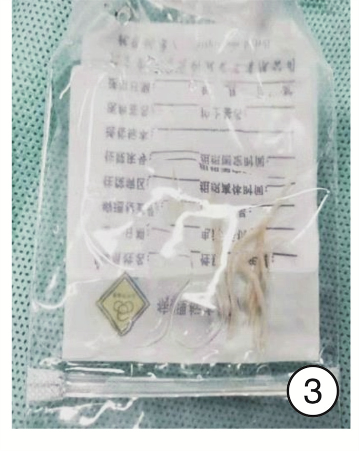
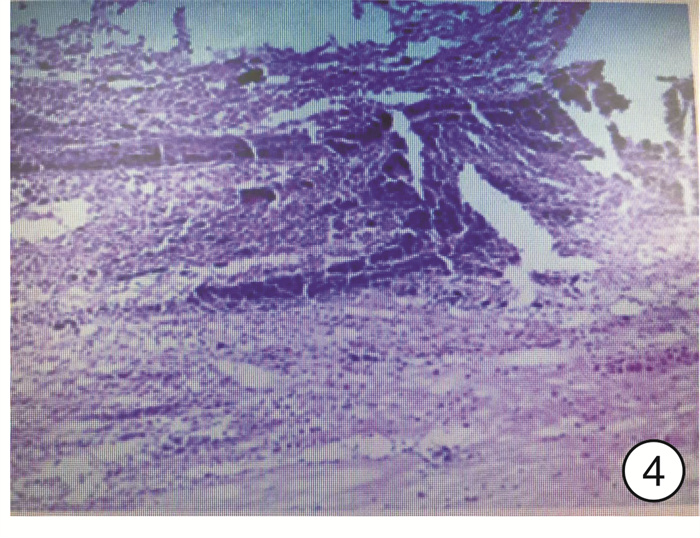
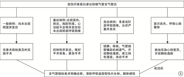
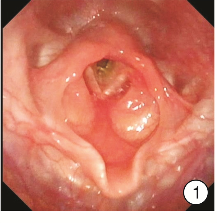
摘要: 目的 探讨硬管支气管镜联合高频通气在小儿急性纤维蛋白性喉气管支气管炎诊治中的作用。方法 回顾性分析7例小儿急性纤维蛋白性喉气管支气管炎患儿的临床资料。7例均应用硬管支气管镜联合高频通气方式行喉及支气管检查术。术中均于声门下、气管、支气管取出大量膜性痂样内生物。结果 6例入院后紧急行手术治疗,痊愈出院; 1例因呼吸衰竭行呼吸机机械通气48 h无好转后行手术取出内生异物,术后终因多器官功能衰竭死亡。7例内生异物病理组织学检查为纤维蛋白性渗出及坏死组织,伴大量炎症细胞浸润。结论 全身麻醉下及时经硬管支气管镜联合高频通气取出堵塞呼吸道的塑型性内生异物是诊断及治疗小儿急性纤维蛋白性喉气管支气管炎的有效方法。一旦疑诊,必须抓紧手术时机,尽快解除呼吸道梗阻,缓解缺氧是减少死亡率的关键。
-
关键词:
- 纤维蛋白性喉气管支气管炎 /
- 支气管镜检查 /
- 喉梗阻 /
- 高频通气 /
- 儿童
Abstract: Objective To explore the role of rigid bronchoscope combined with high frequency ventilation in the diagnosis and treatment of infantile acute fibrinous laryngotracheobronchitis.Method The clinical data of 7 children with acute fibrinous laryngotracheobronchitis were analyzed retrospectively. Laryngology and bronchoscopy were conducted by hard tube bronchoscopy combined with high frequency ventilation in all cases. During the operation, a large quantity of membranous scabs was removed from subglottic area, trachea and bronchus.Result Six cases were treated by emergency operation and cured. One patient was treated with mechanical ventilation for 48 hours because of respiratory failure. Then the operation was performed to remove the endogenous foreign body since no improvement was observed after prolonged ventilation. This patient died of multiple organ failure. The histopathological examination of these 7 cases of endogenous foreign bodies showed fibrinous exudation and necrosis, accompanied by a large quantity of inflammatory cells infiltration.Conclusion Removal of the plastic endogenous foreign bodies which block the respiratory tract by rigid bronchoscope and high frequency ventilation under general anesthesia facilitates the diagnosis and treatment of acute fibrinous laryngotracheobronchitis in pediatric patients. Prompt surgical intervention could relieve the obstruction of respiratory tract, which is crucial to reduce mortality. -

-
表 1 7例急性纤维蛋白渗出性喉气管支气管炎患儿临床资料
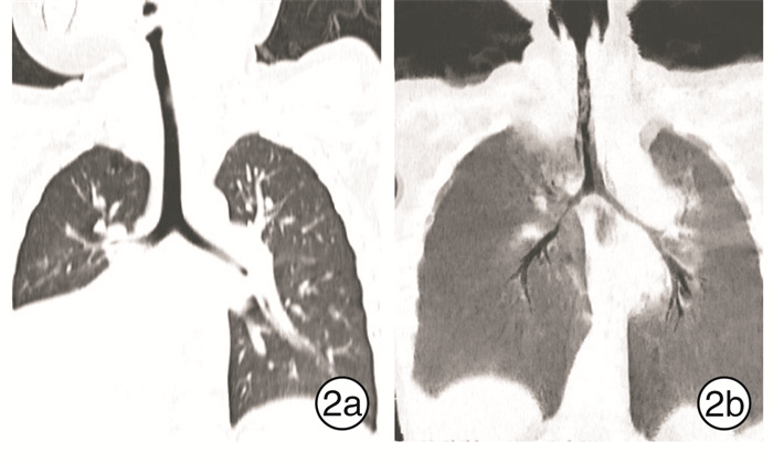
例序 性别 年龄/岁 发病时间/h 呼吸困难程度 影像学检查 手术时机 预后 1 男 1 48 Ⅲ度 右肺上下叶炎症,右侧胸膜增厚 入院后4 h 痊愈 2 男 4.5 24 Ⅳ度 双肺纹理紊乱,纵隔及皮下积气 入院后48 h 死亡 3 男 2 10 Ⅲ度 双肺肺炎,右肺气肿 入院后8 h 痊愈 4 女 1 48 Ⅲ度 双肺肺炎,气道内胸腔入口水平软组织密度影,不除外异物 入院后4 h 痊愈 5 男 2 24 Ⅱ度 右中间段支气管不通畅,可疑异物,右肺中下叶实变 入院后1 h 痊愈 6 男 1 7 d×24 Ⅲ度 右中间段支气管及分支内软组织密度影,右下叶不张,右中叶炎性实变,右侧胸腔积液 入院后8 h 痊愈 7 女 1 24 Ⅱ度 双肺炎性实变 入院后4 h 痊愈 -
[1] 黄选兆, 汪吉宝. 实用耳鼻咽喉科学[M]. 北京: 人民卫生出版社, 2000: 586-586.
[2] Bjomson CL, Johnson DW. Croup[J]. Lancet, 2008, 371(9609): 329-339. doi: 10.1016/S0140-6736(08)60170-1
[3] Rosychuk RJ, Klassen TP, Metes D, et al. Croup presentations to emergency departments in Alberta, Canada: a large population-basedstudy[J]. Pediatr Pulmonol, 2010, 45(1): 83-91. doi: 10.1002/ppul.21162
[4] 刘天夫, 任树北, 朱旭, 等. 支气管镜在小儿急性纤维蛋白性喉气管支气管炎急诊处理中的应用[J]. 临床耳鼻咽喉头颈外科杂志, 2015, 29(16): 1484-1485. https://www.cnki.com.cn/Article/CJFDTOTAL-LCEH201516022.htm
[5] 王双乐, 杨楚, 李创伟, 等. 小儿危重呼吸道阻塞的临床诊断和治疗[J]. 中华耳鼻咽喉头颈外科杂志, 2006, 41(3): 251-254. https://www.cnki.com.cn/Article/CJFDTOTAL-ZHEB200604004.htm
[6] 陈俊宇, 何颜霞. 塑型性支气管炎研究进展[J]. 中华实用儿科临床杂志, 2018, 33(20): 1596-1600. doi: 10.3760/cma.j.issn.2095-428X.2018.20.021
[7] 曾其毅, 刘大波, 罗仁忠, 等. 儿童塑型性支气管炎的诊断与治疗[J]. 中国实用儿科杂志, 2004, 19(2): 81-83. doi: 10.3969/j.issn.1005-2224.2004.02.008
[8] Robinson M, Smiley M, Kotha K, et al. Plastic Bronchitis Treated With Topical Tissue-Type Plasminogen Activator and Cryotherapy[J]. Clin Pediatr(Phila), 2016, 55(12): 1171-1175. doi: 10.1177/0009922815614358
[9] 郭伟, 徐勇胜, 万莉雅, 等. 组织型纤溶酶原激活剂治疗儿童塑型性支气管炎的疗效[J]. 中华实用儿科临床杂志, 2015, 30(16): 1233-1235. doi: 10.3760/cma.j.issn.2095-428X.2015.16.011
[10] Eason DE, Cox K, Moskowitz WB. Aerosolised heparin in the treatment of Fontan-related plastic bronchitis[J]. Cardiol Young, 2014, 24(1): 140-142. doi: 10.1017/S1047951112002089
[11] 杨刚. 纤维支气管镜联合高频喷射通气治疗手术后肺不张38例分析[J]. 安徽医药, 2006, 10(11): 866-866. doi: 10.3969/j.issn.1009-6469.2006.11.043
-


 下载:
下载:
