The distribution of perivascular-resident cells in blood-labyrinth barrier observed with two-photon fluorescence microscope and Imaris deconvolution
-
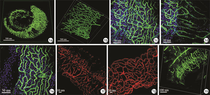
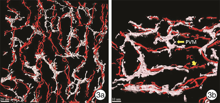
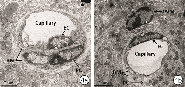
摘要: 目的 明确耳蜗外侧壁血管纹血迷路屏障的细胞构成及分布,分析血管旁固有巨噬细胞(PVM)细胞在血迷路屏障中与周细胞(PC)、内皮细胞的接触关系。方法 分离GFP转基因小鼠(C57BL/6)血管纹铺片,双光子荧光显微镜Z轴逐层扫描并3D重建,观察血迷路屏障中的毛细血管走行。取Balb/c小鼠耳蜗血管纹进行多重免疫荧光染色,经Imaris 3D渲染后观察PVM、PC细胞分布及与毛细血管基膜(BM)的接触模式。电镜下分析血迷路屏障中内皮细胞、PC和PVM细胞的超微结构。结果 血管纹毛细血管走行与其外侧螺旋韧带中的毛细血管网络截然不同,后者平行于蜗轴而前者垂直于蜗轴。血管纹中大量PC细胞紧密缠绕在内皮细胞外侧,与内皮细胞共同包裹在血管BM内; PVM则位于BM外侧。与每个PC细胞仅缠绕单根毛细血管不同,PVM可以分出多个胞质突起而接触多条毛细血管。结论 双光子激光共聚焦荧光显微镜Z轴扫描和Imaris软件3D渲染技术可以清晰显示耳蜗外侧壁血管网立体细微结构,有助于更好地研究血迷路屏障结构和功能。PVM细胞发出多个胞质而与多条毛细血管相接触的分布方式提示可能在血迷路屏障结构中扮演比PC细胞更重要的角色。Abstract: Objective To determine the cell types composing blood-labyrinth barrier in stira vascularis of cochlear lateral wall, analyze the distribution of these composing cells in blood-labyrinth barrier, and to investigate the relationship between perivascular-resident macrophages (PVMs), endothelial cells and pericytes in blood-labyrinth barrier.Method Cochlear lateral wall tissues were harvested from adult GFP-transgenic mice(C57BL/6). Then the isolated whole stria vascularis tissue was scanned at 0.5 um intervals on the Z axis by two-photon confocal microscope and a 3D-structure of stria vascularis was reconstructed to observe the distribution of capillaries in blood-labyrinth barrier. Cochlear stria vascularis isolated from Balb/c mice was stained by mulit-immunofluorescence and then 3D real time deconvolution of stria vascularis was performed by Imaris software to investigate the distribution of PVMs and pericytes, and their contacting with basement membrane of capillaries was also observed. The ultrastructure of endothelial cells, pericytes and PVMs in blood-labyrinth barrier was observed using transmission electron microscope.Result The vessels in stria vascularis are parallel to modiolus, distinct from that in spiral ligament which are perpendicular to modiolus. Numerous pericyts in stria vascularis are ensheathed by a vascular basement membrane shared with endothelial cells and closely attaching to the lateral wall of endothelial cells, while PVMs are located outside basement membrane of capillaries. Unlike pericytes that surround one capillary, PVMs branch to connect with more than one capillary.Conclusion Serial layers on the Z axis scanned by two-photon confocal microscope and a 3D-structure reconstructed by Imaris 3D deconvolution helps to display the micro structure of capillaries in cochlear lateral wall clearly, which could be applied to analyze the 3D structure and function of blood-labyrinth barrier. PVMs in stria vascularis contact with more than one vessel through cytoplasmic processes, suggesting that PVMs may play a more significant role than pericytes in the integrity of blood-labyrinth barrier.
-

-
[1] Shi X. Pathophysiology of the cochlear intrastrial fluid-blood barrier(review)[J]. Hear Res, 2016, 338: 52-63. doi: 10.1016/j.heares.2016.01.010
[2] Kudo T, Wangemann P, Marcus DC. Claudin expression during early postnatal development of the murine cochlea[J]. BMC Physiol, 2018, 18(1): 1-1. doi: 10.1186/s12899-018-0035-1
[3] 潘明金, 张学渊. 豚鼠耳蜗微血管内皮细胞通透性分子调控机制的初步研究[J]. 临床耳鼻咽喉科杂志, 2006, 20(4): 180-183. doi: 10.3969/j.issn.1001-1781.2006.04.014
[4] Le TN, Blakley BW. Mannitol and the blood-labyrinth barrier[J]. J Otolaryngol Head Neck Surg, 2017, 46(1): 66-66. doi: 10.1186/s40463-017-0245-8
[5] Canis M, Bertlich M. Cochlear Capillary Pericytes[J]. Adv Exp Med Biol, 2019, 1122: 115-123.
[6] Liu YH, Zhang ZP, Wang Y, et al. Electrophysiological properties of strial pericytes and the effect of aspirin on pericyte K+ channels[J]. Mol Med Rep, 2018, 17(2): 2861-2868.
[7] 刘艳辉, 王艳萍, 王洋, 等. 豚鼠耳蜗血管纹PC电生理特性的研究[J]. 中华耳鼻咽喉头颈外科杂志, 2016, 51(8): 600-605. doi: 10.3760/cma.j.issn.1673-0860.2016.08.008
[8] Bertlich M, Ihler F, Weiss BG, et al. Cochlear Pericytes Are Capable of Reversibly Decreasing Capillary Diameter In Vivo After Tumor Necrosis Factor Exposure[J]. Otol Neurotol, 2017, 38: e545-e550. doi: 10.1097/MAO.0000000000001523
[9] Jiang Y, Zhang J, Rao Y, et al. Lipopolysaccharide disrupts the cochlear blood-labyrinth barrier by activating perivascularresident macrophages and up-regulating MMP-9[J]. Int J Pediatr Otorhinolaryngol, 2019, 127: 109656. doi: 10.1016/j.ijporl.2019.109656
[10] Zhang W, Zheng J, Meng J, et al. Macrophage migration inhibitory factor knockdown inhibit viability and induce apoptosis of PVM/Ms[J]. Mol Med Rep, 2017, 16(6): 8643-8648. doi: 10.3892/mmr.2017.7684
[11] Neng L, Zhang F, Kachelmeier A, et al. Endothelial cell, pericyte, and perivascular resident macrophage-type melanocyte interactionsregulate cochlear intrastrial fluid-blood barrier permeability[J]. J Assoc Res Otolaryngol, 2013, 14(2): 175-185. doi: 10.1007/s10162-012-0365-9
[12] Zhang J, Chen S, Cai J, et al. Culture media-based selection of endothelial cells, pericytes, and perivascular-resident macrophage-like melanocytes from the young mouse vestibular system[J]. Hear Res, 2017, 345: 10-22. doi: 10.1016/j.heares.2016.12.012
[13] Gamage PPKM, Patel BA, Yeoman MS, et al. Interstitial cell network volume is reduced in the terminal bowel of ageing mice[J]. J Cell Mol Med, 2018, 22(10): 5160-5164. doi: 10.1111/jcmm.13794
[14] Song W, Fhu CW, Ang KH, et al. The fetal mouse metatarsal bone explant as a model of angiogenesis[J]. Nat Protoc, 2015, 10(10): 1459-1473. doi: 10.1038/nprot.2015.097
-


 下载:
下载:


