Analysis of the positive rate of otitis media in 1-12 years old children based on brain MRI
-
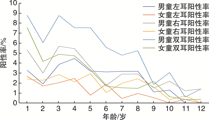
摘要: 目的 通过对儿童颅脑MRI影像提示的中耳炎阳性率进行分析,探讨1~12岁儿童中耳炎的影像学阳性率。方法 收集2014年1月—2020年12月就诊于山东大学附属儿童医院的1~12岁儿童颅脑MRI图像,将MRI扫描野中出现中耳炎症改变定义为阳性,得到中耳炎阳性率,依据不同年龄计算患儿侧别、不同性别之间的患病率,对结果进行分析。结果 12439例患儿中诊断出中耳炎1321例,总阳性率为10.62%,其中男892例,阳性率为67.52%,女429例,阳性率为32.48%,男性阳性率高于女性,并且是女性的1.83倍;男女性患儿阳性率与年龄呈负相关(P < 0.01);男性患儿左耳、右耳及双耳阳性率均高于同龄女性(P < 0.05);中耳炎患儿的左耳和右耳阳性率比较差异无统计学意义(P=0.76)。结论 对中耳炎患儿进行颅脑MRI检查,可明确中耳腔炎症及乳突气房内积液情况。2岁患儿阳性率有陡降趋势,可能是因为乳突气化加速,鼓室内环境改变,鼓室内气房增多,气压发生改变可以抵消由于咽鼓管功能不良导致的负压,使中耳炎发病率降低。Abstract: Objective To investigate the positive imaging rate of otitis media in children aged 1-12 years by analyzing the positive rate of otitis media suggested by cranial magnetic resonance imaging(MRI) images in children.Methods By collecting the brain MRI images of children aged 1-12 in Department of Otolaryngology, Jinan children's Hospital from January 2014 to December 2020, the overall incidence of otitis media and mastoiditis was firstly determined, and then it was divided into 12 age groups according to age, each age group was split into boy and girl groups according to gender, each group was divided into left, right and bilateral groups, with the changes of otitis media and mastoiditis in the scanning field as the positive standard statistical analysis of the results.Results Among 12 439 children in the study, 1321 cases were diagnosed with tympanitis, with a positive rate of 10.62%. Among them, 892 patients were male, with a positive rate of 67.52%, and 429 cases were female, with a positive rate of 32.48%. The positive rate of the male was higher than that of female children, 1.84 times higher than that of female children. The positive momentum in male and female children was negatively correlated with age (P < 0.01). The favorable rates of male children in the left ear, right ear, and both ears were higher than those in female children of the same age(P < 0.05). There was no difference in the positive rate of the left and right ear in children with tympanitis (P=0.76).Conclusion Craniocerebral MRI examination in children with tympanitis can clarify the inflammation of the middle ear cavity and the effusion in the mastoid air chamber. The positive rate of children at two years old showed a steep decline, which may be due to the acceleration of mastoid gasification, the change of tympanic environment, the increase of air chamber in the tympanic room, the evolution of air pressure could offset the negative pressure caused by poor Eustachian tube function, to reduce the incidence of tympanitis.
-
Key words:
- child /
- otitis media /
- magnetic resonance imaging
-

-
[1] Libwea JN, Kobela M, Ndombo PK, et al. The prevalence of otitis media in 2-3 year old Cameroonian children estimated by tympanometry[J]. Int J Pediatr Otorhinolaryngol, 2018, 115: 181-187. doi: 10.1016/j.ijporl.2018.10.007
[2] Bowatte G, Tham R, Perret JL, et al. Air Pollution and Otitis Media in Children: A Systematic Review of Literature[J]. Int J Environ Res Public Health, 2018, 15(2): 257. doi: 10.3390/ijerph15020257
[3] Edmondson-Jones M, Dibbern T, Hultberg M, et al. The effect of pneumococcal conjugate vaccines on otitis media from 2005 to 2013 in children aged ≤ 5 years: a retrospective cohort study in two Swedish regions[J]. Hum Vaccin Immunothe, 2021, 17(2): 517-526. doi: 10.1080/21645515.2020.1775455
[4] van Ingen G, le Clercq CMP, Jaddoe VWV, et al. Identifying distinct trajectories of acute otitis media in children: A prospective cohort study[J]. Clin Otolaryngol, 2021, 46(4): 788-795. doi: 10.1111/coa.13736
[5] 刘娅, 孙建军. 儿童分泌性中耳炎多国指南研读与解析[J]. 临床耳鼻咽喉头颈外科杂志, 2020, 34(12): 1065-1069. doi: 10.13201/j.issn.2096-7993.2020.12.003 https://lceh.cbpt.cnki.net/WKC/WebPublication/paperDigest.aspx?paperID=8018dbcd-50f8-4b55-9a55-f8f2ac2a3b24
[6] Songu M, Islek A, Imre A, et al. Risk factors for otitis media with effusion in children with adenoid hypertrophy[J]. Acta Otorhinolaryngol Ital, 2020, 40(2): 133-137. doi: 10.14639/0392-100X-2456
[7] DeLacy J, Dune T, Macdonald JJ. The social determinants of otitis media in aboriginal children in Australia: are we addressing the primary causes? A systematic content review[J]. BMC Public Health, 2020, 20(1): 492. doi: 10.1186/s12889-020-08570-3
[8] 张鹏, 王延飞, 蒲章杰, 等. 山东省滨州市儿童分泌性中耳炎流行病学调查[J]. 中华耳科学杂志, 2009, 7(4): 367-370. doi: 10.3969/j.issn.1672-2922.2009.04.024
[9] 王智楠, 陈平, 徐忠强, 等. 武汉市部分幼儿园儿童分泌性中耳炎患病率调查[J]. 临床耳鼻咽喉头颈外科杂志, 2009, 23(22): 1036-1037, 1043. https://lceh.cbpt.cnki.net/WKC/WebPublication/paperDigest.aspx?paperID=9f184191-c3a7-4007-a762-d1518e281795
[10] 唐红燕, 胡瑞丹, 李庆, 等. 成都市2~7岁儿童分泌性中耳炎患病现状调查[J]. 听力学及言语疾病杂志, 2019, 27(1): 83-84. https://www.cnki.com.cn/Article/CJFDTOTAL-TLXJ201901021.htm
[11] 唐志辉, 虞玮翔, 顾家铭, 等. 中国香港与西方儿童分泌性中耳炎发病率的比较[J]. 中华耳鼻咽喉科杂志, 2004, 39(7): 51-54. https://www.cnki.com.cn/Article/CJFDTOTAL-ZHEB200407019.htm
[12] 王进东, 张再兴, 孙静涛, 等. 唐山地区2008~2013年儿童急性中耳炎流行病学调查[J]. 中国妇幼保健, 2015, 30(6): 939-941. https://www.cnki.com.cn/Article/CJFDTOTAL-ZFYB201506048.htm
[13] Varsak YK, Gül Z, Eryılmaz MA, et al. Prevalence of otitis media with effusion among school age children in rural parts of Konya province, Turkey[J]. Kulak Burun Bogaz Ihtis Derg, 2015, 25(4): 200-204. doi: 10.5606/kbbihtisas.2015.01979
[14] Mark A, Matharu V, Dowswell G, et al. The point prevalence of otitis media with effusion in secondary school children in Pokhara, Nepal: a cross-sectional study[J]. Int J Pediatr Otorhinolaryngol, 2013, 77(9): 1523-1529.
[15] Maharjan M, Bhandari S, Singh I, et al. Prevalence of otitis media in school going children in Eastern Nepal[J]. Kathmandu Univ Med J(KUMJ), 2006, 4(4): 479-482.
[16] Gultekin E, Develioǧlu ON, Yener M, et al. Prevalence and risk factors for persistent otitis media with effusion in primary school children in Istanbul, Turkey[J]. Auris Nasus Larynx, 2010, 37(2): 145-149.
[17] 刘玉红, 苏法仁. 分泌性中耳炎的相关发病机制及治疗研究[J]. 中华耳科学杂志, 2018, 16(2): 234-238. https://www.cnki.com.cn/Article/CJFDTOTAL-ZHER201802021.htm
-


 下载:
下载:
