Nasal endoscopy assisting combined with transoral approach for resection of carcinoma of the palate invading nasal cavity and sinuses
-
摘要: 目的 探讨鼻内镜辅助联合经口入路切除侵犯鼻腔鼻窦的上腭恶性肿瘤的应用价值。方法 回顾性分析21例原发于硬腭及上颌牙槽的上腭恶性肿瘤患者病历资料,术前鼻内镜、CT和MRI检查,所有患者原发灶肿瘤均不同程度侵犯鼻腔鼻窦或颅底区域,均采用鼻内镜辅助联合经口入路切除原发灶,术后根据病理类型和临床分期辅以放疗或同步放化疗,随访并统计分析术后并发症、肿瘤全切率、肿瘤局部控制率和5年生存率。结果 所有患者均成功经此联合入路实施肿瘤切除,18例获得肿瘤整块切除,切缘阴性。中位随访期60个月,所有患者术后鼻腔通气功能良好,无溢泪,总体原发灶局部控制率85.7%,总体5年生存率76.2%。结论 鼻内镜辅助联合经口入路是切除侵犯鼻腔鼻窦的上腭恶性肿瘤的一种有效方法,有利于肿瘤整块切除,控制原发灶;有利于鼻腔鼻窦功能保留,符合功能性外科原则。Abstract: Objective To explore the value of nasal endoscopy assisting combined with transoral approach in resection of the carcinoma of the palate with the nasal cavity and sinuses invaded.Methods A retrospective analysis of 21 patients with a primary malignant tumors of the palate was performed. Preoperative nasal endoscopy and CT and MRI scan showed that the primary tumors invading the nasal cavity and sinuses in all patients or skull base with varying degrees. All patients were treated by nasal endoscopic assisting combined with transoral approach. Postoprational adjuvant radiotherapy or concurrent chemoradiotherapy was performed according to pathological types and clinical stage. Postoperative complications, all-tumor resection rate, local control rate and 5-year survival rate were analyzed statistically.Results The combined approach was successfully performed in all patients. En bloc resection was carried out in 18 patients by this combined approach and surgical margins were free of carcinoma. The median follow-up period was 60 months. All patients had good nasal ventilation function and no epiphora in postoperation, and the overall local control rate of primary site was 85.7%, overall 5-year survival rate was 76.2%.Conclusion Nasal endoscopy assisting combined with transoral approach is an effective method for the resection of palate malignant tumors invading the nasal cavity and sinuses. It is convenient for en bloc resection and local control of primary lesions. It is beneficial to preserve the function of nasal cavity and sinuses, which is in line with the principle of functional surgery.
-
Key words:
- nasal endoscopy /
- combined approach /
- carcinoma of palate
-

-
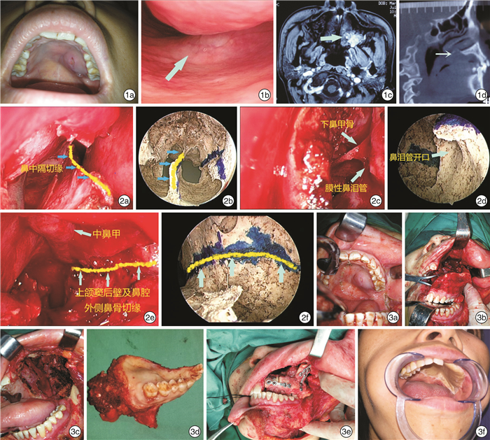
图 1 术前检查肿瘤外观及影像学表现 1a:口腔检查肿瘤外观;1b:鼻内镜检查示肿瘤突入鼻底;1c:磁共振扫描显示肿瘤累及上颌窦后外侧壁、翼突下份;1d:CT扫描显示肿瘤突破硬腭进入鼻底; 图2 术中鼻内镜处理标志线与干性颅骨标本对照图 2a:鼻内镜下处理鼻中隔切缘(箭头所示);2b:相应鼻内镜下干性头颅标本示意图;2c:鼻内镜下处理鼻腔外侧壁切缘,保留膜性鼻泪管;2d:相应鼻内镜下干性头颅标本示意图,显示骨性鼻泪管鼻腔开口(箭头所示);2e:鼻内镜下处理上颌窦后壁及鼻腔外侧壁后份切缘;2f:相应鼻内镜下干性头颅标本示意图; 图3 术中及术后随访 3a:口腔标记肿瘤切缘;3b:经口截骨后肿瘤标本松动;3c:肿瘤切除后创面;3d:切除的肿瘤标本;3e:游离腓骨瓣就位;3f:术后4年复查口内像。
-
[1] Morris LG, Patel SG, Shah JP, et al. High rates of regional failure in squamous cell carcinoma of the hard palate and maxillary alveolus[J]. Head Neck, 2011, 33(6): 824-830. doi: 10.1002/hed.21547
[2] Aydil U, Kızıl Y, Bakkal FK, et al. Neoplasms of the hard palate[J]. J Oral Maxillofac Surg, 2014, 72(3): 619-626. doi: 10.1016/j.joms.2013.08.019
[3] Warnakulasuriya S. Global epidemiology of oral and oropharyngeal cancer[J]. Oral Oncol, 2009, 45(4/5): 309-316.
[4] 李慧, 马士崟, 张明洁, 等. 鼻内镜辅助下开放径路联合放疗治疗中晚期上颌窦恶性肿瘤的疗效分析[J]. 临床耳鼻咽喉头颈外科杂志, 2017, 31(14): 1078-1081. https://www.cnki.com.cn/Article/CJFDTOTAL-LCEH201714006.htm
[5] Ozawa H, Sekimizu M, Saito S, et al. Endoscopic Endonasal Management of Pterygopalatine Fossa Tumors[J]. J Craniofac Surg, 2021, 32(5): e454-e457.
[6] 刘婷婷, 王学海, 蔡晓岚, 等. 鼻内镜下口-鼻联合入路切除鼻底累及硬腭巨大肿瘤两例[J]. 山东大学耳鼻喉眼学报, 2018, 32(3): 108-111. https://www.cnki.com.cn/Article/CJFDTOTAL-SDYU201803024.htm
[7] Piastro K, Chen T, Khatiwala RV, et al. Endoscopic endonasal resection of cavernous hemangioma of the palate[J]. Otolaryngol Case Reports, 2017, 5: 13-15. doi: 10.1016/j.xocr.2017.08.001
[8] Yang X, Song X, Chu W, et al. Clinicopathological Characteristics and Outcome Predictors in Squamous Cell Carcinoma of the Maxillary Gingiva and Hard Palate[J]. J Oral Maxillofac Surg, 2015, 73(7): 1429-1436. doi: 10.1016/j.joms.2014.12.034
[9] Yang Z, Deng R, Sun G, et al. Cervical metastases from squamous cell carcinoma of hard palate and maxillary alveolus: a retrospective study of 10 years[J]. Head Neck, 2014, 36(7): 969-975. doi: 10.1002/hed.23398
[10] Wang TC, Hua CH, Lin CC, et al. Risk factors affect the survival outcome of hard palatal and maxillary alveolus squamous cell carcinoma: 10-year review in a tertiary referral center[J]. Oral Surg Oral Med Oral Pathol Oral Radiol Endod, 2010, 110(1): 11-17. doi: 10.1016/j.tripleo.2009.11.035
[11] Hakim SG, Steller D, Sieg P, et al. Clinical course and survival in patients with squamous cell carcinoma of the maxillary alveolus and hard palate: Results from a single-center prospective cohort[J]. J Craniomaxillofac Surg, 2020, 48(1): 111-116. doi: 10.1016/j.jcms.2019.12.008
-


 下载:
下载: