
-
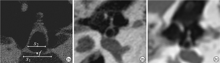
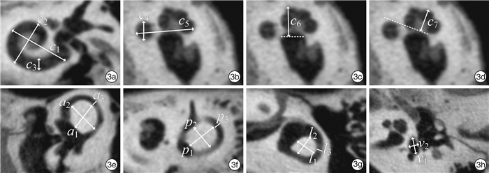
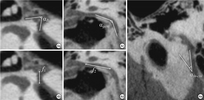
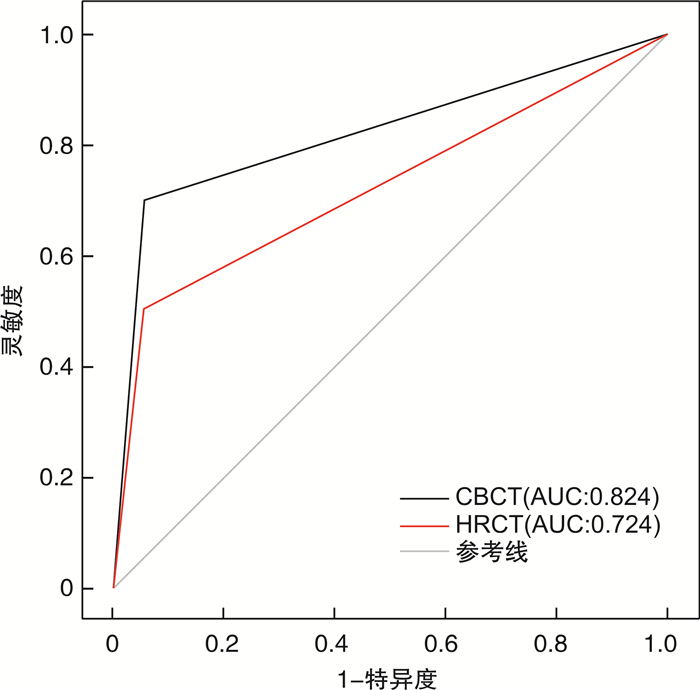
摘要: 目的 探究锥形束计算机断层扫描(CBCT)在颞骨细微结构测量的可行性及正常值范围。方法 15例福尔马林浸泡固定的人类尸头标本分别行CBCT、高分辨率CT、Micro CT扫描,对包括听骨链、耳蜗、半规管及面神经等在内的中内耳结构进行形态学测量,比较三种扫描方法所测结果间差异。结果 三种不同CT扫描所测参数组间对比除镫骨底板厚度(P < 0.01)及耳蜗底周管径(P < 0.01)外,余差异均无统计学意义。CBCT对面神经骨管缺损具有较好的诊断价值。结论 CBCT扫描时间短,辐射剂量小,成像质量高,能较准确地显示颞骨内各精细结构的形态特点,是耳科影像学的可靠评估方法。
-
关键词:
- 锥形束计算机断层扫描 /
- 多平面重建 /
- 颞骨
Abstract: Objective To identified the feasibility and normal range of cone beam computer tomography(CBCT) in the measurement of temporal bone.Methods 15 formalin fixed human cadaver head specimens were scanned by CBCT, high resolution CT, and Micro CT, respectively. Morphological parameter measurements of the middle and inner ear structures including ossicular chain, cochlea, semicircular canal and facial nerve were performed, and the results measured by the three scanning methods were compared.Results None of the parameters measured by the three scanning methods were statistically significant except the thickness of stapes footplate(P < 0.01) and the diameter of cochlear basal turn(P < 0.01). CBCT was superior in detecting facial nerve bony canal dehiscence.Conclusion CBCT has the advantages of short scanning time, low radiation dose and high resolution. It can accurately display the morphological characteristics of the temporal bone structures, and is a reliable evaluation method for otological surgery. -

-
表 1 不同CT扫描方法测得锤、砧、镫骨及面神经变异情况 x±s
部位 Micro CT CBCT HRCT P 锤骨柄长度/mm 4.40±0.22 4.47±0.37 4.38±0.27 >0.05 锤骨头长径/mm 3.33±0.20 3.33±0.14 3.38±0.18 >0.05 锤骨头宽径/mm 2.26±0.19 2.30±0.12 2.32±0.14 >0.05 砧骨长脚长度/mm 3.90±0.25 3.86±0.15 3.88±0.17 >0.05 砧骨短脚长度/mm 2.32±0.21 2.38±0.21 2.40±0.24 >0.05 砧骨长短脚夹角/(°) 116.72±2.63 116.63±1.16 116.18±1.69 >0.05 砧骨长脚下缘至底板距离/mm 3.66±0.24 3.70±0.15 3.70±0.10 >0.05 镫骨底板厚度/mm 0.29±0.05 0.38±0.04 0.44±0.04 < 0.01 镫骨底板长度/mm 2.57±0.25 2.61±0.16 2.57±0.17 >0.05 镫骨前后弓距离/mm 1.78±0.25 1.73±0.12 1.72±0.10 >0.05 砧镫关节角度/(°) 94.79±2.89 95.32±2.04 94.75±1.81 >0.05 面神经前膝角/mm 83.94±4.41 83.98±1.61 83.23±1.91 >0.05 面神经迷路段长/mm 2.80±0.24 2.75±0.24 2.83±0.27 >0.05 面神经鼓室段长/mm 10.27±0.25 10.12±0.32 10.25±0.23 >0.05 面神经后膝角/(°) 126.47±3.31 126.91±0.95 126.81±1.07 >0.05 与鼓索神经夹角/(°) 26.77±1.89 26.21±1.57 26.23±1.09 >0.05  下载: 导出CSV
表 2 不同CT扫描方法测得耳蜗、前庭、半规管变异情况
mm,x±s 部位 Micro CT CBCT HRCT P 耳蜗底周长径 8.85±0.55 8.86±0.54 8.85±0.49 >0.05 耳蜗底周宽径 6.64±0.43 6.52±0.32 6.57±0.38 >0.05 耳蜗底周管径 2.02±0.15 1.95±0.18 1.66±0.15 < 0.01 耳蜗底周上半周宽度 7.14±0.28 7.02±0.26 7.09±0.40 >0.05 耳蜗底转高度 2.07±0.15 2.07±0.10 2.04±0.14 >0.05 耳蜗中周高度 2.99±0.21 2.95±0.11 2.97±0.17 >0.05 耳蜗高度 3.68±0.19 3.64±0.18 3.63±0.23 >0.05 前庭长径 4.45±0.35 4.34±0.26 4.36±0.20 >0.05 前庭宽径 2.77±0.43 2.73±0.36 2.72±0.35 >0.05 上半规管高度 6.00±0.18 6.13±0.39 6.14±0.38 >0.05 上半规管宽度 5.88±0.32 5.92±0.18 5.86±0.32 >0.05 上半规管内径 0.92±0.12 0.96±0.07 0.98±0.05 >0.05 后半规管高度 5.45±0.65 5.47±0.54 5.43±0.68 >0.05 后半规管宽度 4.80±0.51 4.93±0.45 4.99±0.48 >0.05 后半规管内径 1.13±0.13 1.09±0.07 1.10±0.11 >0.05 外半规管高度 3.74±0.61 3.83±0.49 3.87±0.32 >0.05 外半规管宽度 3.42±0.30 3.43±0.61 3.49±0.28 >0.05 外半规管内径 1.17±0.12 1.16±0.12 1.14±0.06 >0.05
下载: 导出CSV
表 3 CBCT和HRCT对面神经骨管完整性的判断
CT 敏感性/% 特异性/% 约登指数 Kappa值 P CBCT 70.0 94.7 0.647 0.680 >0.05 HRCT 50.0 94.7 0.447 0.485 >0.05
下载: 导出CSV
-
[1] Nasseh I, Al-Rawi W. Cone Beam Computed Tomography[J]. Dent Clin North Am, 2018, 62(3): 361-391. doi: 10.1016/j.cden.2018.03.002
[2] Walliczek-Dworschak U, Diogo I, Strack L et al. Indications of cone beam CT in head and neck imaging in children[J]. Acta Otorhinolaryngol Ital, 2017, 37(4): 270-275. doi: 10.14639/0392-100X-1219
[3] Stutzki M, Jahns E, Mandapathil MM, et al. Indications of cone beam CT in head and neck imaging[J]. Acta Otolaryngol, 2015, 135(12): 1337-1343. doi: 10.3109/00016489.2015.1076172
[4] Patel S, Dawood A, Ford TP, et al. The potential applications of cone beam computed tomography in the management of endodontic problems[J]. Int Endod J, 2007, 40(10): 818-830. doi: 10.1111/j.1365-2591.2007.01299.x
[5] Redfors YD, Grondahl HG, Hellgren J, et al. Otosclerosis: anatomy and pathology in the temporal bone assessed by multi-slice and cone-beam CT[J]. Otol Neurotol, 2012, 33(6): 922-927. doi: 10.1097/MAO.0b013e318259b38c
[6] Razafindranaly V, Truy E, Pialat JB, et al. Cone Beam CT Versus Multislice CT: Radiologic Diagnostic Agreement in the Postoperative Assessment of Cochlear Implantation[J]. Otol Neurotol, 2016, 37(9): 1246-1254. doi: 10.1097/MAO.0000000000001165
[7] Komori M, Yanagihara N, Hyodo J, et al. Position of TORP on the stapes footplate assessed with cone beam computed tomography[J]. Otol Neurotol, 2012, 33(8): 1353-1356. doi: 10.1097/MAO.0b013e31826a5260
[8] 宋跃帅, 龚树生. 应用锥形束CT评估人工耳蜗植入后电极形态的研究[J]. 临床耳鼻咽喉头颈外科杂志, 2018, 32(18): 1371-1373. https://www.cnki.com.cn/Article/CJFDTOTAL-LCEH201818002.htm
[9] 黄健健, 夏巍, 唐翔龙, 等. 锥形束CT在人工耳蜗植入中的研究进展[J]. 临床耳鼻咽喉头颈外科杂志, 2021, 35(6): 567-572. https://www.cnki.com.cn/Article/CJFDTOTAL-LCEH202106021.htm
[10] 巩武贤, 巩若箴, 赵斌, 等. 正常人耳蜗标准化HRCT径线测量[J]. 医学影像学杂志, 2018, 28(8): 1254-1257. https://www.cnki.com.cn/Article/CJFDTOTAL-XYXZ201808009.htm
[11] 张征宇, 王争, 赵鹏飞, 等. 锥形束CT观察成人离体面神经管膝状神经窝影像学解剖[J]. 中国医学影像技术, 2021, 37(2): 284-288. https://www.cnki.com.cn/Article/CJFDTOTAL-ZYXX202102037.htm
[12] 阮士栋, 巩武贤, 樊兆民, 等. 慢性中耳炎患者听骨链功能状态的HRCT评价[J]. 医学影像学杂志, 2016, 26(5): 791-794, 803. https://www.cnki.com.cn/Article/CJFDTOTAL-XYXZ201605009.htm
[13] 宋跃帅, 龚树生. 内耳及相关解剖结构的教学方法和经验[J]. 中华耳科学杂志, 2020, 10(1): 208-213. doi: 10.3969/j.issn.1672-2922.2020.01.037
[14] Gupta R, Bartling SH, Basu SK et al. Experimental flat-panel high-spatial-resolution volume CT of the temporal bone[J]. AJNR Am J Neuroradiol, 2004, 25(8): 1417-1424.
[15] Penninger RT, Tavassolie TS, Carey JP. Cone-beam volumetric tomography for applications in the temporal bone[J]. Otol Neurotol, 2011, 32(3): 453-460. doi: 10.1097/MAO.0b013e31820d962c
[16] Yamashita K, Hiwatashi A, Togao O, et al. Ultrahigh-resolution CT scan of the temporal bone[J]. Eur Arch Otorhinolaryngol 2018, 275(11): 2797-2803. doi: 10.1007/s00405-018-5101-6
[17] Curthoys IS, Uzun-Coruhlu H, Wong CC, et al. The configuration and attachment of the utricular and saccular maculae to the temporal bone. New evidence from microtomography-CT studies of the membranous labyrinth[J]. Ann N Y Acad Sci, 2009, 1164: 13-18. doi: 10.1111/j.1749-6632.2008.03729.x
[18] Zou J, Hannula M, Lehto K, et al. X-ray microtomographic confirmation of the reliability of CBCT in identifying the scalar location of cochlear implant electrode after round window insertion[J]. Hear Res, 2015, 326: 59-65. doi: 10.1016/j.heares.2015.04.005
[19] Arai Y, Tammisalo E, Iwai K, et al. Development of a compact computed tomographic apparatus for dental use[J]. Dentomaxillofac Radiol, 1999, 28(4): 245-248. doi: 10.1038/sj.dmfr.4600448
[20] Small BW. Cone beam computed tomography[J]. Gen Dent 2007, 55(3): 179-181.
[21] Dahmani-Causse M, Marx M, Deguine O, et al. Morphologic examination of the temporal bone by cone beam computed tomography: comparison with multislice helical computed tomography[J]. Eur Ann Otorhinolaryngol Head Neck Dis, 2011, 128(5): 230-235. doi: 10.1016/j.anorl.2011.02.016
[22] Dalchow CV, Weber AL, Yanagihara N, et al. Digital volume tomography: radiologic examinations of the temporal bone[J]. AJR Am J Roentgenol, 2006, 186(2): 416-423. doi: 10.2214/AJR.04.1353
[23] Lata S, Mohanty SK, Vinay S, et al. Is Cone Beam Computed Tomography(CBCT)a Potential Imaging Tool in ENT Practice? A Cross-Sectional Survey Among ENT Surgeons in the State of Odisha, India[J]. Indian J Otolaryngol Head Neck Surg, 2018, 70(1): 130-136. doi: 10.1007/s12070-017-1168-4
[24] 唐作华, 钱雯, 宋济昌, 等. 耳部正常解剖结构的CT数据测量[J]. 临床放射学杂志, 2004, 23(7): 566-570. doi: 10.3969/j.issn.1001-9324.2004.07.006
[25] Gluth MB, Cohen MA, Friedland PL, et al. Malleostapedotomy prosthesis size and shape: key measurements from a temporal bone study[J]. Otol Neurotol, 2012, 33(4): 518-522. doi: 10.1097/MAO.0b013e31824b7882
[26] 徐宁, 巩若箴, 巩武贤. 多层螺旋CTMPR技术对镫骨结构的显示及相关数据测量[J]. 医学影像学杂志, 2014, 24(2): 193-196. https://www.cnki.com.cn/Article/CJFDTOTAL-XYXZ201402009.htm
[27] 顾于燕, 任冬冬, 韩朝. 中国成人正常实体镫骨精确测量及临床意义[J]. 中国眼耳鼻喉科杂志, 2017, 17(1): 16-18. https://www.cnki.com.cn/Article/CJFDTOTAL-YRBH201701010.htm
[28] 柴麦娥, 郝楷, 刘书平. 砧骨的测量及临床应用[J]. 解剖学杂志, 1991, 14(4): 384-386. https://www.cnki.com.cn/Article/CJFDTOTAL-JPXZ199104040.htm
[29] Kaftan H, Bohme A, Martin H. Is the prosthesis length in malleostapedotomy for otosclerosis revision surgery predictable?[J]. Otol Neurotol, 2014, 35(7): 1150-1155. doi: 10.1097/MAO.0000000000000448
[30] Gosselin E, Elblidi A, Alhabib SF, et al. Predictable prosthesis length on a high-resolution CT scan before a stapedotomy[J]. Eur Arch Otorhinolaryngol, 2018, 275(9): 2219-2226. doi: 10.1007/s00405-018-5075-4
[31] 杨颖, 夏明, 边志强, 等. 高分辨CT正常内耳解剖结构的测量研究[J]. 山东大学学报(医学版), 2008, 46(11): 1075-1079. https://www.cnki.com.cn/Article/CJFDTOTAL-SDYB200811017.htm
[32] 曾令延, 魏文洲, 李茂进, 等. 正常人内耳结构的MR测量[J]. 医学影像学杂志, 2006, 16(3): 226-229. https://www.cnki.com.cn/Article/CJFDTOTAL-XYXZ200603003.htm
[33] Sudhoff H, Gehl HB, Boga E, et al. Stapes Prosthesis Length: One Size Fits All?[J]. Audiol Neurootol, 2019, 24(1): 1-7. doi: 10.1159/000494915
[34] Zou J, Lahelma J, Arnisalo A, et al. Clinically relevant human temporal bone measurements using novel high-resolution cone-beam CT[J]. J Otol, 2017, 12(1): 9-17. doi: 10.1016/j.joto.2017.01.002
[35] 马晓波, 赵守琴, 李洁, 等. 正常耳颞骨内面神经形态分析[J]. 中国耳鼻咽喉头颈外科, 2015, 22(6): 287-289. https://www.cnki.com.cn/Article/CJFDTOTAL-EBYT201506010.htm
[36] McManus LJ, Dawes PJ, Stringer MD. Clinical anatomy of the chorda tympani: a systematic review[J]. J Laryngol Otol, 2011, 125(11): 1101-1108. doi: 10.1017/S0022215111001873
[37] Revesz P, Piski Z, Burian A, et al. Delayed Facial Paralysis following Uneventful KTP Laser Stapedotomy: Two Case Reports and a Review of the Literature[J]. Case Rep Med, 2014, 2014: 971362.
[38] Zhang Z, Yin H, Wang Z, et al. Imaging re-evaluation of the tympanic segment of the facial nerve canal using cone-beam computed tomography compared with multi-slice computed tomography[J]. Eur Arch Otorhinolaryngol, 2019, 276(7): 1933-1941. doi: 10.1007/s00405-019-05419-3
-

图(6)
表(3)
计量
- 文章访问数: 2233
- PDF下载数: 485
- 施引文献: 0