Applied value of color Doppler flow imaging in diagnosis of congenital subglottic haemangioma in infant
-
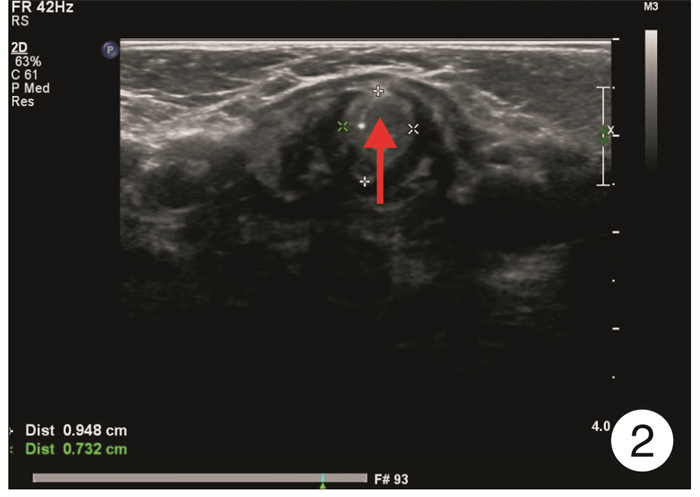
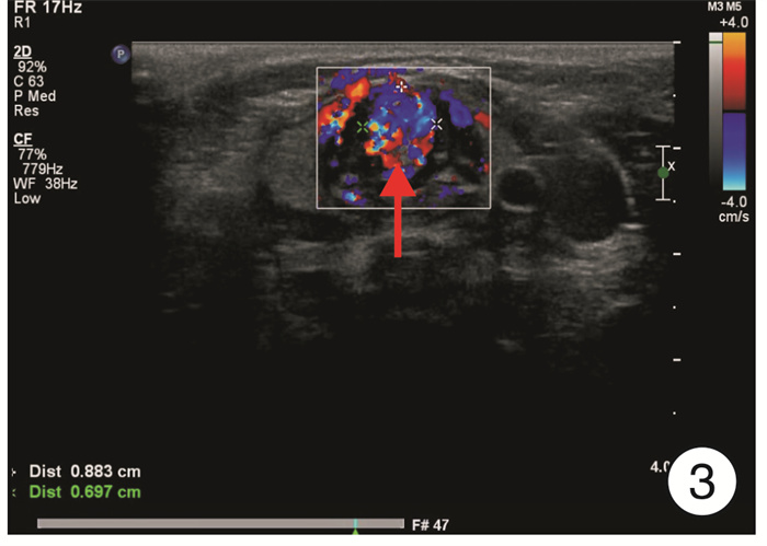
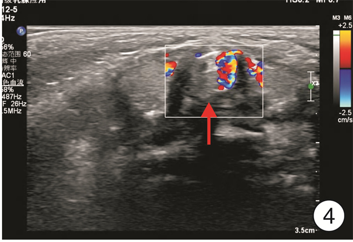
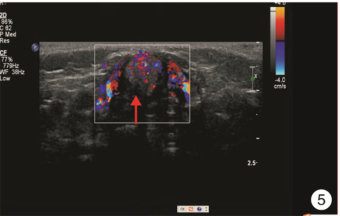
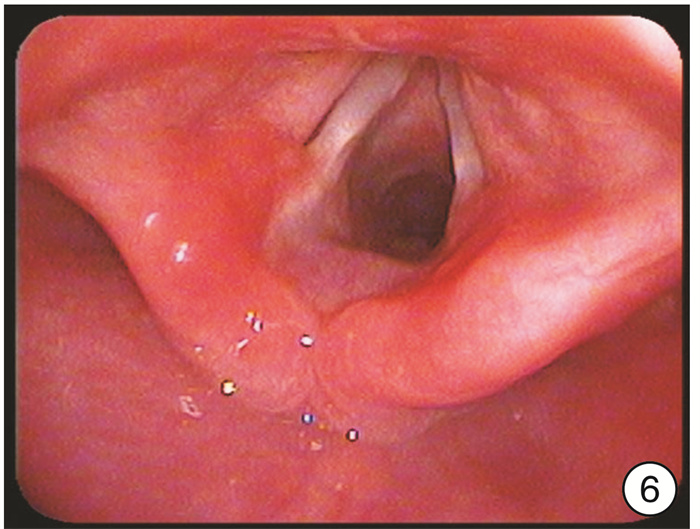
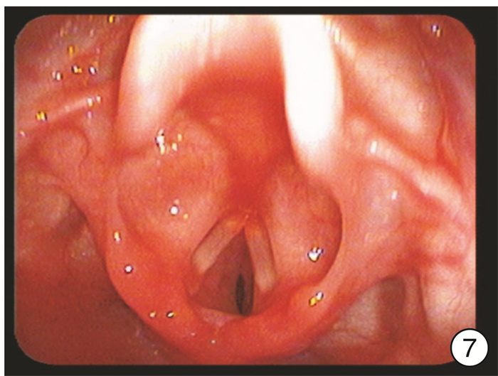
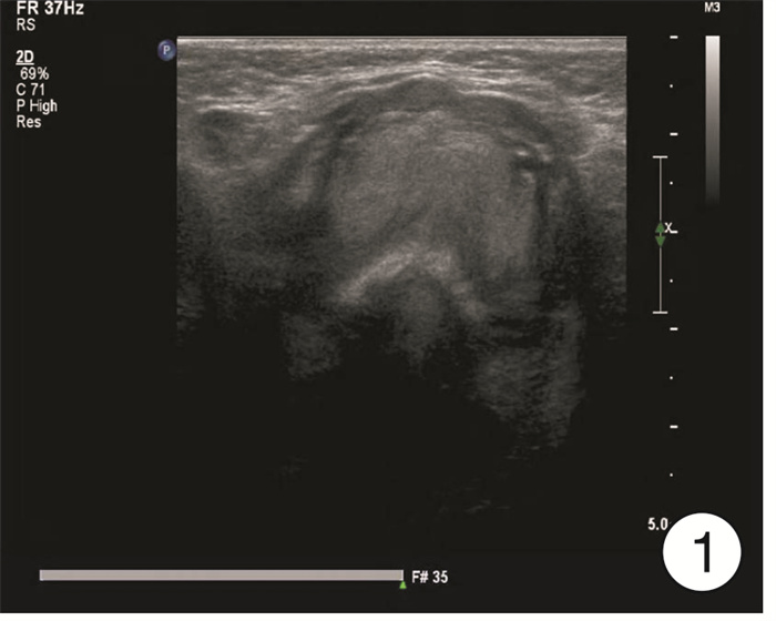
摘要: 目的 探讨彩色多普勒血流显像(CDFI)技术在婴幼儿先天性声门下血管瘤(CSH)诊断中的价值。 方法 回顾性分析行喉部CDFI检查的18例CSH患儿资料,观察正常喉部及CSH的图像特点及瘤体外形、大小、血流特点。18例患儿均给予口服普萘洛尔治疗,分别于治疗1周、1个月、3个月后复查喉部CDFI。 结果 CDFI可清晰显示18例声门下血管瘤的位置、形态、大小、范围以及与气道、周围组织的关系。声门下血管瘤CDFI图像显示瘤体呈团块状或结节状,内见丰富血流信号或者斑片状血流信号。血管瘤位于声门下右侧壁6例,声门下左侧壁8例,双侧4例。 结论 CDFI技术可以应用于声门下血管瘤的诊断,在显示其大小、范围、与气道关系等方面具有优势,特别是在后期的治疗随访中更简便快捷。Abstract: Objective To investigate the value of color Doppler flow imaging(CDFI) in the diagnosis of congenital subglottic hemangioma(CSH) in infants. Methods The data of 18 children with CSH who underwent laryngeal CDFI examination were collected and analyzed retrospectively, and compared with those who underwent laryngeal ultrasound examination at the same time. The shape, size, blood flow characteristics of the tumor and its relationship with airway were observed. Eighteen cases were treated with propranolol orally. CDFI of larynx was reexamined after 1 week, 1 month and 3 months of treatment. Results CDFI could clearly show the location, shape, size and range of CSH in 18 cases, as well as the relationship with airway and surrounding tissues. CDFI images of CSH showed that the tumor was massive or nodular with abundant or patchy blood flow signals. Hemangioma was found in 6 cases on the right side, 8 cases on the left side, and 4 cases on both sides. Conclusion CDFI can be used in the diagnosis of subglottic hemangioma. It has advantages in displaying its size, scope and relationship with airway, especially in the later treatment and follow-up.
-
Key words:
- infant /
- congenital subglottic hemangioma /
- glottis /
- color Doppler flow imaging
-

-
表 1 18例CSH患儿初次就诊超声检查结果
例序 性别 年龄 位置 大小/cm CDFI 1 男 18天10小时 左侧 0.3×0.2×0.4 斑片状血流信号 2 男 3个月17天 左侧 0.3×0.3×0.4 斑片状血流信号 3 男 1天8小时 左侧 0.4×0.3×0.3 斑片状血流信号 4 男 1个月17天 声门下 0.8×0.8×0.4 丰富血流信号 5 男 2个月3天 左侧 0.5×0.3×0.4 丰富血流信号 6 男 1个月19天 右侧 0.6×0.4×0.5 丰富血流信号 7 女 1岁6个月 右侧 1.0×0.8×0.4 丰富血流信号 8 女 1岁8个月 声门下 1.0×1.0×0.7 丰富血流信号 9 女 2个月28天 右侧 0.8×0.9×0.8 丰富血流信号 10 女 6个月24天 声门下 0.3×0.3×0.7 丰富血流信号 11 女 1岁4月 右侧 0.9×0.7×0.6 丰富血流信号 12 女 5个月21天 左侧 0.5×0.3×0.3 斑片状血流信号 13 女 2个月10天 左侧 0.4×0.4×0.5 斑片状血流信号 14 女 1个月3天 左侧 0.4×0.3×0.4 丰富血流信号 15 女 9天10小时 右侧 0.2×0.3×0.4 丰富血流信号 16 女 4个月11天 声门下 0.5×0.5×0.4 丰富血流信号 17 女 4天3小时 左侧 0.3×0.4×0.4 丰富血流信号 18 女 20天9小时 右侧 0.3×0.5×0.4 丰富血流信号 -
[1] 王桂香, 张丰珍, 张亚梅, 等. 普萘洛尔治疗婴幼儿声门下血管瘤临床疗效及远期随访观察[J]. 中国耳鼻咽喉头颈外科, 2020, 27(8): 464-468. https://www.cnki.com.cn/Article/CJFDTOTAL-EBYT202008011.htm
[2] 高胜利, 陈彦球, 邹宇, 等. 低温等离子消融术治疗婴幼儿声门下血管瘤临床观察[J]. 临床耳鼻咽喉头颈外科杂志, 2013, 27(12): 656-659. https://www.cnki.com.cn/Article/CJFDTOTAL-LCEH201312012.htm
[3] 辛渊, 马淑巍, 陈洁. 窄带成像内镜在小儿声门下血管瘤中的应用[J]. 中华耳鼻咽喉头颈外科杂志, 2016, 51(12): 956.
[4] Matsuzawa-Kinomura Y, Ozeki M, Otsuka H, et al. Neonatal dysphonia caused by subglottic infantile hemangioma[J]. Pediatr Int, 2017, 59(8): 935-936. doi: 10.1111/ped.13308
[5] Kumar P, Kaushal D, Garg PK, et al. Subglottic hemangioma masquerading as croup and treated successfully with oral propranolol[J]. Lung India, 2019, 36(3): 233-235.
[6] Onder SS, Gergin O, Karabulut B. A Life Threatening Subglottic and Mediastinal Hemangioma in an Infant[J]. J Craniofac Surg, 2019, 30(5): e402-e404. doi: 10.1097/SCS.0000000000005340
[7] 张忠晓, 刘霞, 马静, 等. 婴幼儿声门下血管瘤24例支气管镜诊断及疗效评估[J]. 中华实用儿科临床杂志, 2015, 30(16): 1241-1244. doi: 10.3760/cma.j.issn.2095-428X.2015.16.013
[8] 胡迪, 孙记航, 路春兰, 等. 64层螺旋CT血管造影三维及多平面重组在婴儿声门下血管瘤的应用[J]. 临床放射学杂志, 2012, 31(4): 550-552. https://www.cnki.com.cn/Article/CJFDTOTAL-LCFS201204026.htm
[9] Rossler L, Rothoeft T, Teig N, et al. Ultrasound and colour Doppler in infantile subglottic haemangioma[J]. Pediatr Radiol, 2011, 41(11): 1421-1428. doi: 10.1007/s00247-011-2213-1
-


 下载:
下载:
