
-
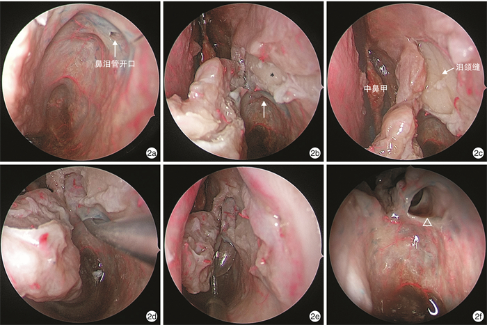
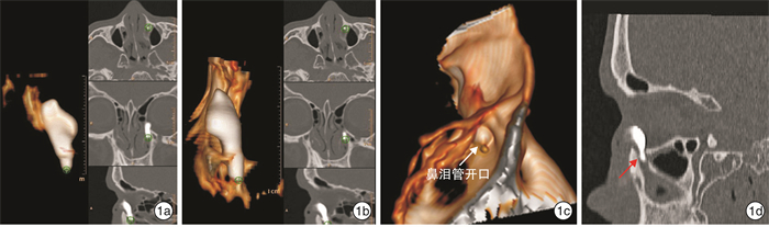
摘要: 目的 探讨鼻内镜下功能性鼻泪管解压术治疗慢性泪囊炎的可行性。方法 收集13例22侧慢性泪囊炎患者, 给予30%碘海醇注射液进行泪囊造影, 造影剂从泪小管返流停止推药, 立即进行泪道CT扫描。应用Sinuses Trachea Ⅰ软件重建泪道及其周围结构三维可视图; 应用该软件对泪囊、鼻泪管模拟"切削", 去除腹内侧泪囊-鼻泪管1/2~3/4周进行骨管减压, 暴露膜性鼻泪管。对10例成人冷冻尸头进行CT扫描, 模拟鼻泪管解压手术, 磨去鼻泪管的骨管, 暴露膜性鼻泪管, 并对膜性鼻泪管进行球囊导管扩张。结果 ① 泪道造影发现, 泪道阻塞多发生在鼻泪管段, 占研究病例的72.7%(16/22)。②解剖显示鼻泪管外侧壁由上颌骨的泪沟构成, 内壁由泪骨降突构成。③尸头模拟解压鼻泪管骨管, 暴露膜性鼻泪管。④球囊导管扩张膜性鼻泪管, 冲洗泪道通畅。结论 鼻内镜下鼻泪管解压术治疗慢性泪囊炎保留了膜性泪道完整性和虹吸功能, 可减少因切开泪囊肉芽增生或瘢痕形成所致泪道再次阻塞等并发症的发生。Abstract: Objective To investigate the feasibility of endoscopic nasolacrimal decompression for chronic dacryocystitis.Method 22 patients with chronic dacryocystitis hospitalized at Longgang ENT hospital were participated in this study. An injection of 30% iohexol was administered to conduct lacrimal sac angiography. The injection was stopped when the agent reflux from the lacrimal duct, and a computed tomography(CT) scan of the lacrimal duct was performed immediately. Sinuses Trachea Isoftware was used to reconstruct a three-dimensional(3D) view of the lacrimal passage and its surrounding structures. The software was used to simulate the "cutting" of the lacrimal sac and nasolacrimal duct; the lacrimal sac and nasal lacrimal duct were removed after 1/2-3/4 circumferences to decompress the passage and expose the membranous nasolacrimal duct. CT scans were performed on ten adult frozen cadaveric heads, and the nasolacrimal duct decompression operation was simulated. Then, the bone of the nasolacrimal duct was removed, membranous nasolacrimal duct was exposed, and the capsular nasolacrimal duct was dilated.Result ① The lacrimal angiography study revealed that lacrimal duct obstruction occurred in the nasolacrimal duct segment, accounting for 72.7%(16/22) of the study cases. ② The anatomical examination showed that the outer sidewall of the nasolacrimal duct was composed of the tear groove of the maxilla, and the inner wall was composed of the descending process of the lacrimal bone. ③ In cadaveric heads, decompression of the osseous nasolacrimal duct was performed, exposing the membranous nasolacrimal duct. ④ A balloon catheter could dilate the membranous nasolacrimal duct and allow the lacrimal passage to be flushed.Conclusion Endoscopic nasolacrimal decompression preserves the integrity of the lacrimal duct, allows drainage of the lacrimal duct, and avoids obstruction of the lacrimal duct by preventing lacrimal granulation.
-

-
[1] 訾魁然. 鼻内窥镜下鼻腔泪囊造孔术联合泪道置管治疗慢性泪囊炎临床疗效观察[J]. 实用中西医结合临床, 2019, 19(4): 113-115. https://www.cnki.com.cn/Article/CJFDTOTAL-SZXL201904059.htm
[2] Wu S, Xu T, Fan B, et al. Endoscopic dacryocystorhinostomy with an otologic T-type ventilation tube in repeated revision cases[J]. BMC Ophthalmol, 2017, 17(1): 138. doi: 10.1186/s12886-017-0539-7
[3] 李晓晖, 刘智献, 王鹏, 等. 鼻泪管解压术治疗慢性泪囊炎的影像学研究[J]. 临床耳鼻咽喉头颈外科杂志, 2017, 31(4): 290-292. https://www.cnki.com.cn/Article/CJFDTOTAL-LCEH201704011.htm
[4] Ali MJ, Paulsen F. Etiopathogenesis of Primary Acquired Nasolacrimal Duct Obstruction: What We Know and What We Need to Know[J]. Ophthalmic Plast Reconstr Surg, 2019, 35(5): 426-433. doi: 10.1097/IOP.0000000000001310
[5] 杨培新, 朱雪妙, 吴创奇, 等. 鼻内镜下泪囊鼻腔吻合联合丝裂霉素C治疗慢性泪囊炎19例临床分析[J]. 中国耳鼻咽喉颅底外科杂志, 2015, 21(5): 420-421. https://www.cnki.com.cn/Article/CJFDTOTAL-ZEBY201505026.htm
[6] 徐菁, 焦秦, 蔡昌枰. 内镜下泪囊鼻腔吻合置管术治疗慢性泪囊炎疗效[J]. 中华眼外伤职业眼病杂志, 2019, 41(3): 172-175. https://www.cnki.com.cn/Article/CJFDTOTAL-LCYZ201904031.htm
[7] Hiremath R, Satyamurthy KV, Kulkarni S, et al. Powered Endoscopic Dacryocystorhinostomy: Raising the Bar[J]. Delhi J Ophthalmol, 2019, 29(4): 44-47.
-
 下载:
下载:
图(2)
计量
- 文章访问数: 891
- PDF下载数: 547
- 施引文献: 0