-
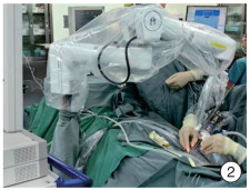
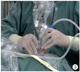
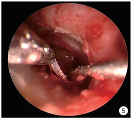
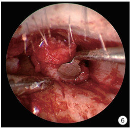
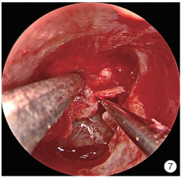
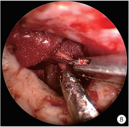
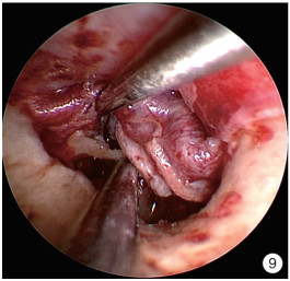
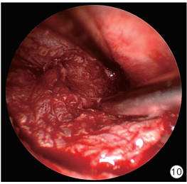
摘要: 目的 探讨支撑内镜技术辅助Ⅰ型鼓室成形术的手术效果及其安全性。 方法 回顾性分析2022年11月—2023年9月在北京协和医院耳鼻咽喉科接受Ⅰ型鼓室成形术的16例患者的临床资料,其中传统耳内镜手术组8例,支撑内镜手术组8例,分析手术流程,观察支撑内镜下操作的完成情况,同时记录手术持续时间、主要步骤耗时、擦拭镜头频率、围术期并发症及术后听力改善情况,并进行统计学分析。 结果 支撑内镜技术实现了耳内镜手术中实时吸除渗血,一手牵拉组织时另一手进行分离,精细去除鼓膜内侧钙化斑,修剪外耳道皮瓣,稳定分离锤骨柄和鼓膜,有张力地复位皮肤软骨膜瓣等操作。支撑内镜组的平均手术持续时间、外耳道皮瓣制作时间和皮肤软骨膜瓣复位时间较对照组减少,平均擦拭镜头频率较对照组明显下降。2组患者术后听力改善情况差异无统计学意义,术后均未发生感染或是因鼓膜再穿孔需要进行二次手术。 结论 支撑内镜技术实现了内镜下双手操作及单双手便捷切换的需求,完成了许多传统内镜手术无法完成的操作,解决了既往术中单手操作及图像不稳定等问题,平均手术时间较传统耳内镜手术缩短,术中擦拭镜头频率较传统耳内镜手术明显下降,具有缩短学习曲线的潜在价值。Abstract: Objective To investigate the surgical outcomes and safety of the follower arm endoscope holder in assisting type Ⅰ tympanoplasty. Methods The clinical data of 16 patients who underwent type Ⅰ tympanoplasty at the Department of Otorhinolaryngology, Peking Union Medical College Hospital, from November 2022 to September 2023 were retrospectively analyzed, among which 8 cases were operated by traditional otoscopy and 8 cases were operated by supported endoscopy.The surgical procedure was analyzed and the completion of supported endoscopic operation was observed, while the duration of the operation, the time consumed by the main steps, the frequency of wiping the lenses, the perioperative complications, and the improvement of the postoperative hearing were recorded and statistically analyzed. Results Supporting endoscopic technology achieved real-time suction of bleeding, simultaneous traction and separation of tissues, precise removal of calcified spots on the inner side of the eardrum, trimming of the external auditory canal flap, stable separation of the handle of the malleus and the eardrum, and tensioned repositioning of the skin-cartilage flap. The average duration of surgery, time for external auditory canal flap preparation, and time for repositioning the skin-cartilage flap were reduced in the supporting endoscopic surgery group compared to the control group. The average lens wiping frequency was significantly lower in the supporting endoscopic surgery group compared to the control group. There was no statistically significant difference in postoperative hearing improvement between the two groups, and no infections or the need for secondary surgery due to eardrum re-perforation occurred postoperatively. Conclusion Supported endoscopy technology realizes the need for endoscopic two-handed operation and convenient switching between one and two hands, accomplishes many operations that cannot be done by traditional endoscopic surgery, solves the problems of previous intraoperative one-handed operation and image instability, shortens the average operation time compared with traditional otoscopic surgery, and decreases the frequency of intraoperative wiping of the lens significantly compared with traditional otoscopic surgery, which is potentially worthwhile in terms of shortening the learning curve.
-

-
表 1 支撑内镜组患者的临床资料
例序 性别 年龄/岁 手术持续时间/min 内镜操作时间/min 擦拭频率/次/h 擦拭次数 皮瓣制作时间/min 皮瓣复位时间/min 外耳道填塞时间/min 1 女 33 70 57.2 2.1 2 14.0 11.0 8.7 2 男 51 93 64.1 4.7 5 19.9 10.6 5.3 3 男 63 121 105.3 4.6 8 20.6 16.3 4.8 4 男 53 80 49.0 0.0 0 17.6 - 5.9 5 女 70 87 65.1 0.9 1 26.2 5.0 10.3 6 女 49 112 97.0 0.6 1 14.6 9.8 11.6 7 女 58 101 69.3 4.3 5 18.8 9.1 5.1 8 女 47 82 55.0 1.1 1 20.4 6.4 6.9 均值 93.3 70.3 2.3 19.0 9.7 7.3 手术持续时间:麻醉单上记录的手术开始至手术结束的时间;内镜操作时间:指在内镜下记录到外耳道局部麻醉至耳甲腔填塞碘仿纱条的时间,需减去游离及修剪耳屏软骨的时间;擦拭频率:术中擦拭耳内镜镜头的频率;擦拭次数:术中擦拭耳内镜镜头的频率;皮瓣制作时间:纵行切开外耳道皮肤至掀起外耳道皮瓣开始探查鼓室的时间间隔;皮瓣复位时间:耳屏软骨及软骨膜进入内镜视野下至外耳道开始填塞明胶海绵的时间;外耳道填塞时间:外耳道填塞明胶海绵的时间。 表 2 传统内镜手术组患者的临床资料
例序 性别 年龄/岁 手术持续时间/min 内镜操作时间/min 擦拭频率/次/h 擦拭次数 皮瓣制作时间/min 皮瓣复位时间/min 外耳道填塞时间/min 1 女 31 100 64.3 5.6 6 21.7 4.6 3.8 2 女 52 68 53.2 12.4 11 23.7 6.0 3.4 3 女 52 84 56.8 4.2 4 19.5 7.8 4.1 4 男 55 135 122.8 15.6 32 36.4 10.2 10.3 5 女 58 133 105.9 18.1 32 19.0 10.5 10.4 6 女 33 79 45.7 2.6 2 18.3 - 3.3 7 女 52 138 124.1 10.2 21 20.3 30.1 7.0 8 女 58 76 58.4 15.4 15 17.7 9.9 5.3 均值 101.6 78.9 10.5 22.1 11.3 6.0 -
[1] 刘业军, 赵亚会. 耳内镜与手术显微镜下鼓膜修补术临床疗效的Meta分析[J/OL]. 中国耳鼻咽喉头颈外科, 2019, 26(8): 449-454.
[2] Elnahal KB, Hassan MA, Maarouf AM. Comparison of endoscope-assisted and microscope-assisted type Ⅰ tympanoplasty; a systematic review and meta-analysis[J]. Eur Arch Otorhinolaryngol, 2023, 15.
[3] Khan MM, Parab SR. Novel Concept of Attaching Endoscope Holder to Microscope for Two Handed Endoscopic Tympanoplasty[J]. Indian J Otolaryngol Head Neck Surg, 2016, 68(2): 230-240. doi: 10.1007/s12070-015-0916-6
[4] Rosen M, Ponsky J. Minimally invasive surgery[J]. Endoscopy, 2001, 33(4): 358-366. doi: 10.1055/s-2001-13689
[5] Goh HK, Ng YH, Teo DT. Minimally invasive surgery for head and neck cancer [J]. Lancet Oncol, 2010, 11(3): 281-286. doi: 10.1016/S1470-2045(09)70379-1
[6] Briner HR, Simmen D, Jones N. Endoscopic sinus surgery: advantages of the bimanual technique[J]. Am J Rhinol, 2005, 19(3): 269-273. doi: 10.1177/194589240501900310
[7] Arnholt JL, Mair EA. A 'third hand' for endoscopic skull base surgery[J]. Laryngoscope, 2002, 112(12): 2244-2249. doi: 10.1097/00005537-200212000-00021
[8] May M, Hoffmann DF, Sobol SM. Video endoscopic sinus surgery: a two-handed technique[J]. Laryngoscope, 1990, 100(4): 430-432. doi: 10.1288/00005537-199004000-00021
[9] Castelnuovo P, Pistochini A, Locatelli D. Different surgical approaches to the sellar region: focusing on the "two nostrils four hands technique"[J]. Rhinology, 2006, 44(1): 2-7.
[10] Fischer M, Gröbner C, Dietz A, et al. Klinischer Einsatz eines Endoskop-Manipulator-Systems in der endo-und transnasalen Chirurgie[First clinical experiences with an endoscope manipulator system in endo-and transnasal surgery][J]. Laryngorhinootologie, 2012, 91(8): 487-493. doi: 10.1055/s-0032-1309053
[11] Hintschich CA, Fischer R, Seebauer C, et al. A third hand to the surgeon: the use of an endoscope holding arm in endonasal sinus surgery and well beyond[J]. Eur Arch Otorhinolaryngol, 2022, 279(4): 1891-1898. doi: 10.1007/s00405-021-06935-x
[12] Paraskevopoulos D, Roth J, Constantini S. Endoscope Holders in Cranial Neurosurgery: Part Ⅰ-Technology, Trends, and Implications[J]. World Neurosurg, 2016, 89: 343-354. doi: 10.1016/j.wneu.2016.01.052
-

| 引用本文: | 殷悦, 赵勇, 陈晗林, 等. 支撑内镜技术在Ⅰ型鼓室成形术中的应用[J]. 临床耳鼻咽喉头颈外科杂志, 2024, 38(2): 112-116. doi: 10.13201/j.issn.2096-7993.2024.02.006 |
| Citation: | YIN Yue, ZHAO Yong, CHEN Hanlin, et al. Application of the follower arm endoscope holder in type i tympanoplasty[J]. J Clin Otorhinolaryngol Head Neck Surg, 2024, 38(2): 112-116. doi: 10.13201/j.issn.2096-7993.2024.02.006 |
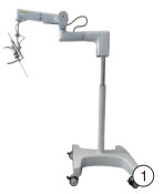
- Figure 1.
- Figure 2.
- Figure 3.
- Figure 4.
- Figure 5.
- Figure 6.
- Figure 7.
- Figure 8.
- Figure 9.
- Figure 10.
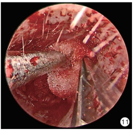
- Figure 11.


 下载:
下载:
