
Application of U-Net network in automatic image segmentation of adenoid and airway of nasopharynx
-
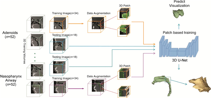
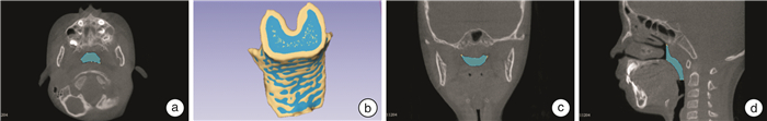
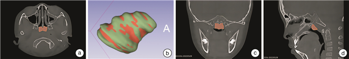
摘要: 目的 探讨基于U-Net网络的深度学习模型对儿童腺样体及鼻咽气道的全自动图像分割效果。方法 2021年3月-2022年3月在深圳大学总医院耳鼻咽喉头颈外科因睡眠打鼾或张口呼吸进行锥形束计算机断层扫描(CBCT)检查的患儿240例,选取其中52例进行鼻咽部和腺样体人工标注,再由深度学习模型训练与验证。将模型应用于剩余188例数据后,比较所有240例数据常规二维指标及深度学习三维指标间的差异。结果 对于52例建模以及训练数据集,深度学习预测结果与人工标注结果差异均无统计学意义(P>0.05),模型评价指标鼻咽气道容积的均交并比为(86.32±0.54)%;相似系数为(92.91±0.23)%;准确度为(95.92±0.25)%;精准度为(91.93±0.14)%;腺样体体积的均交并比为(86.28±0.61)%;相似系数为(92.88±0.17)%;准确度为(95.90±0.29)%;精准度为(92.30±0.23)%。240例不同年龄段患儿二维指标A/N和三维指标AV/(AV+NAV)之间均呈正相关性(P < 0.05),9~14岁的相关系数达0.74。结论 基于U-Net网络的深度学习模型对儿童腺样体及鼻咽气道全自动图像分割效果良好,为今后进一步研究导致OSA的腺样体肥大的三维诊断标准提供有利的大数据计算模型。
-
关键词:
- U-Net网络 /
- 全自动图像分割 /
- 腺样体 /
- 鼻咽气道 /
- 锥形束计算机断层扫描
Abstract: Objective To explore the effect of fully automatic image segmentation of adenoid and nasopharyngeal airway by deep learning model based on U-Net network.Methods From March 2021 to March 2022, 240 children underwent cone beam computed tomography(CBCT) in the Department of Otolaryngology, Head and Neck Surgery, General Hospital of Shenzhen University. 52 of them were selected for manual labeling of nasopharynx airway and adenoid, and then were trained and verified by the deep learning model. After applying the model to the remaining data, compare the differences between conventional two-dimensional indicators and deep learning three-dimensional indicators in 240 datasets.Results For the 52 cases of modeling and training data sets, there was no significant difference between the prediction results of deep learning and the manual labeling results of doctors(P>0.05). The model evaluation index of nasopharyngeal airway volume: Mean Intersection over Union(MIOU) s (86.32±0.54)%; Dice Similarity Coefficient(DSC): (92.91±0.23)%; Accuracy: (95.92±0.25)%; Precision: (91.93±0.14)%; and the model evaluation index of Adenoid volume: MIOU: (86.28±0.61)%; DSC: (92.88±0.17)%; Accuracy: (95.90±0.29)%; Precision: (92.30±0.23)%. There was a positive correlation between the two-dimensional index A/N and the three-dimensional index AV/(AV+NAV) in 240 children of different age groups(P < 0.05), and the correlation coefficient of 9-13 years old was 0.74.Conclusion The deep learning model based on U-Net network has a good effect on the automatic image segmentation of adenoid and nasopharynx airway, and has high application value. The model has a certain generalization ability. -

-
表 1 患儿一般资料
X±S 特征 2~5岁(n=103) 6~9岁(n=106) 10~14岁(n=31) 性别(男/女) 52/51 62/44 20/11 年龄/岁 4.45±0.91 7.85±1.15 11.38±1.06 身高/cm 109.95±7.96 129.79±11.59 148.32±13.19 体重/kg 18.62±4.33 27.49±9.04 44.87±14.03 BMI/(kg/m2) 15.25±2.10 15.95±2.70 19.88±3.93  下载: 导出CSV
表 2 深度学习分割与医生人工标注比较
X±S 特征 医生人工标注 深度学习分割 t P 腺样体体积/mm3 2 387.30±572.82 2 647.00±591.80 1.231 0.43 鼻咽气道体积/mm3 3 336.00±664.00 3 877.18±871.54 0.490 0.81
下载: 导出CSV
表 3 模型评估指标
X±S 特征 均交并比/% 相似系数/% 准确度/% 精准度/% 腺样体体积 86.28±0.61 92.88±0.17 95.90±0.29 92.30±0.23 鼻咽部气道体积 86.32±0.54 92.91±0.23 95.92±0.25 91.93±0.14
下载: 导出CSV
表 4 CBCT的二维指标、三维指标深度学习机器测量结果
X±S 特征 2~5岁组(n=103) 6~9岁组(n=106) 10~14岁组(n=31) 二维指标 咽部气道宽度/mm 4.44±1.87 6.81±2.56 8.41±3.52 腺样体厚度/mm 15.73±2.75 15.14±2.87 15.39±3.09 鼻咽腔宽度/mm 19.97±2.23 21.93±1.96 23.60±2.77 腺样体厚度/鼻咽腔宽度 0.78±0.09 0.69±0.11 0.66±0.13 三维指标 腺样体体积/mm3 2 142.20±759.07 2 420.96±920.61 2 566.32±797.11 鼻咽气道体积/mm3 3 163.86±1 262.76 4 141.02±1 395.45 4 778.77±2 263.27 腺样体体积/鼻咽腔体积+腺样体体积 0.35±0.13 0.32±0.15 0.30±0.16
下载: 导出CSV
-
[1] 师炎敏, 裴晓婷, 李润涛, 等. CT容积模型分析儿童腺样体大小与鼻咽腔容积的相关性[J]. 中国医学影像技术, 2020, 36(3): 377-381. https://www.cnki.com.cn/Article/CJFDTOTAL-ZYXX202003016.htm
[2] 杨文麒, 张亚琼, 郭靖晗, 等. 儿童阻塞性睡眠呼吸暂停低通气综合征上气道形态的锥形束CT研究[J]. 口腔疾病防治, 2022, 30(11): 792-797. https://www.cnki.com.cn/Article/CJFDTOTAL-GDYB202211005.htm
[3] 徐光宪, 冯春, 马飞. 基于UNet的医学图像分割综述[J/OL]. 计算机科学与探索, 2023: 1-18.
[4] Fujioka M, Young LW, Girdany BR. Radiographic evaluation of adenoidal size in children: adenoidal-nasopharyngeal ratio[J]. AJR Am J Roentgenol, 1979, 133(3): 401-404. doi: 10.2214/ajr.133.3.401
[5] Anwar SM, Majid M, Qayyum A, et al. Medical Image Analysis using Convolutional Neural Networks: A Review[J]. J Med Syst, 2018, 42(11): 226. doi: 10.1007/s10916-018-1088-1
[6] Huang SY, Hsu WL, Hsu RJ, et al. Fully Convolutional Network for the Semantic Segmentation of Medical Images: A Survey[J]. Diagnostics (Basel), 2022, 12(11): 2765. doi: 10.3390/diagnostics12112765
[7] Leonardi R, Lo Giudice A, Farronato M, et al. Fully automatic segmentation of sinonasal cavity and pharyngeal airway based on convolutional neural networks[J]. Am J Orthod Dentofacial Orthop, 2021, 159(6): 824-835. doi: 10.1016/j.ajodo.2020.05.017
-

图(4)
表(4)
计量
- 文章访问数: 803
- PDF下载数: 163
- 施引文献: 0