
Diagnostic value of computed tomographic cisternography and magnetic resonance hydrography in cerebrospinal fluid rhinorrhea
-
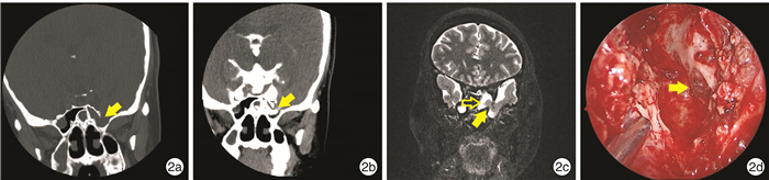
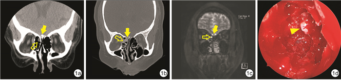
摘要: 目的评估术前CT脑池造影(CTC)与头颅核磁共振水成像(MRH)对脑脊液鼻漏诊断的准确性。方法回顾性分析天津市环湖医院耳鼻咽喉头颈外科2016年10月—2022年1月收治的确诊为脑脊液鼻漏且同时完成术前HRCT、CTC以及MRH检查的38例患者资料,结合术中发现的漏口位置比较其与术前影像学检查结果的一致性。结果38例脑脊液鼻漏患者中,HRCT、CTC、头颅MRH检查发现漏口的比率分别为31.58%、89.47%和60.53%,CTC的准确率高于MRH(χ2=8.49,P=0.007),MRH的准确率高于HRCT(χ2=6.41,P=0.01)。按脑脊液鼻漏漏口的发生部位进行亚组分析,脑脊液鼻漏漏口位于筛窦者中,CTC(80.95% vs.14.29%,P < 0.01)与MRH(52.38% vs.14.29%,P=0.02)对漏口定位的准确率均显著高于HRCT,然而CTC与MRH之间的定位准确率差异无统计学意义(P=0.10); 在脑脊液鼻漏漏口位于蝶窦者中,CTC对漏口定位的准确率显著高于MRH(100.00% vs.61.54%,P=0.04)与HRCT(100.00% vs.53.85%,P < 0.01),然而MRH与HRCT之间的定位准确率差异无统计学意义(P=1.00); 在脑脊液鼻漏漏口位于额窦者中,CTC、MRH以及HRCT之间对漏口定位的准确率差异均无统计学意义。在HRCT未能发现脑脊液鼻漏漏口的26例患者中,CTC与MRH对漏口定位的准确率分别为84.62%和57.69%,差异无统计学意义(χ2=4.59,P=0.06)。结论影像学检查技术的综合运用对脑脊液鼻漏的正确治疗及预后评价有重要的指导意义。CTC与MRH可提高对脑脊液鼻漏漏口定位诊断的准确率。Abstract: ObjectiveTo evaluate the accuracy of preoperative computed tomographic cisternography(CTC) and magnetic resonance hydrography(MRH)in the diagnosis of cerebrospinal fluid(CSF) rhinorrhea.MethodsRetrospective analysis was made on the data of 38 patients diagnosed as cerebrospinal rhinorrhea who completed preoperative HRCT, CTC and MRH examinations in the Department of Otolaryngology Head and Neck Surgery of Tianjin Huanhu Hospital from October 2016 to January 2022. The diagnostic accuracy of preoperative imaging examinations was compared according to the leak location found during operation.ResultsAmong all the 38 cases with CSF rhinorrhea, the detection rates of HRCT, CTC, and cranial MRH were 31.58%, 89.47%, and 60.53%, respectively. The accuracy of CTC was significantlyhigher than that of MRH (χ2=8.49, P=0.007), and the accuracy of MRH was significantly higher than that of HRCT (χ2=6.41, P=0.01). Subgroup analyses were performed according to the precise positioning of cerebrospinal fluid rhinorrhea leakage. In patients with cerebrospinal fluid rhinorrhea located in ethmoid sinus, the accuracy of CTC(80.95% vs. 14.29%, P < 0.01)and MRH(52.38% vs. 14.29%, P=0.02)were significantly higher than that of HRCT, but the accuracy but the difference between CTC and MRH between CTC and MRH was not statistically significant (P=0.10). The accuracy of CTC was significantly higher than that of MRH (100.00% vs. 61.54%, P=0.04) and HRCT (100.00% vs. 53.85%, P < 0.01) in patients with CSF rhinorrhea located in sphenoid sinus. However, there was no significant difference in the accuracy between MRH and HRCT (P=1.00). However, There was no significant difference in the accuracy of CTC, MRH and HRCT in patients with cerebrospinal fluid rhinorrhea located in frontal sinus. The accuracy of CTC and MRH was 84.62% and 57.69% respectively in 26 patients who failed to detect a CSF rhinorrhea by HRCT, and the difference was not statistically significant(χ2=4.59, P=0.06).ConclusionThe comprehensive application of the multiple imaging methods has important guiding significance for the accurate treatment and prognosis evaluation. CTC and MRH could improve the accuracy of the localization diagnosis of cerebrospinal fluid rhinorrhea.
-

-
表 1 HRCT、CTC以及MRH检查对不同部位的脑脊液鼻漏漏口定位的准确率比较
部位 HRCT CTC MRH χ2 HRCT vs.CTC vs.MRH HRCT vs.CTC HRCT vs.MRH CTC vs.MRH 筛窦 与术中发现一致/例 3 17 11 18.80 1) 18.71 2) 6.86 2) 3.86 与术中发现不一致/例 18 4 10 准确率/% 14.29 80.95 52.38 蝶窦 与术中发现一致/例 7 13 8 7.85 1) 7.80 2) 0.16 6.19 2) 与术中发现不一致/例 6 0 5 准确率/% 53.85 100.00 61.54 额窦 与术中发现一致/例 2 4 4 4.80 2.67 2.67 - 与术中发现不一致/例 2 0 0 准确率/% 50.00 100.00 100.00 三组比较,1)P < 0.05; 两组比较,2)P < 0.05。  下载: 导出CSV
-
[1] Caballero N, Bhalla V, Stankiewicz JA, et al. Effect of lumbar drain placement on recurrence of cerebrospinal rhinorrhea after endoscopic repair[J]. Int Forum Allergy Rhinol, 2012, 2(3): 222-226. doi: 10.1002/alr.21023
[2] 魏晓明, 张帆, 薛祎腾, 等. CT脑池造影在脑脊液鼻漏定位中的应用[J]. 中国微侵袭神经外科杂志, 2019, 24(12): 547-551. https://www.cnki.com.cn/Article/CJFDTOTAL-ZWQX201912010.htm
[3] Du HS, Ma GS, Ma JJ. Surgical Repair and Detection of Cerebrospinal Fluid Rhinorrhea using Magnetic Resonance Cisternography and Skull Base Coronal Thin-section Computed Tomography Scan[J]. Chin Med J(Engl), 2016, 129(16): 2005-2006.
[4] Goel G, Ravishankar S, Jayakumar PN, et al. Intrathecal gadolinium-enhanced magnetic resonance cisternography in cerebrospinal fluid rhinorrhea: road ahead?[J]. J Neurotrauma, 2007, 24(10): 1570-1575. doi: 10.1089/neu.2007.0326
[5] Parmar H, Gujar S, Shah G, et al. Imaging of the anterior skull base[J]. Neuroimaging Clin N Am, 2009, 19(3): 427-439. doi: 10.1016/j.nic.2009.06.011
[6] Alonso RC, de la Peña MJ, Caicoya AG, et al. Spontaneous skull base meningoencephaloceles and cerebrospinal fluid fistulas[J]. Radiographics, 2013, 33(2): 553-570. doi: 10.1148/rg.332125028
[7] 蔡芳宇, 董栋, 赵玉林. 鼻窦及颅底气化程度与自发性脑脊液鼻漏的相关性分析[J]. 临床耳鼻咽喉头颈外科杂志, 2021, 35(3): 252-255. https://www.cnki.com.cn/Article/CJFDTOTAL-LCEH202103013.htm
[8] Lloyd KM, DelGaudio JM, Hudgins PA. Imaging of skull base cerebrospinal fluid leaks in adults[J]. Radiology, 2008, 248(3): 725-736. doi: 10.1148/radiol.2483070362
[9] Oakley GM, Alt JA, Schlosser RJ, et al. Diagnosis of cerebrospinal fluid rhinorrhea: an evidence-based review with recommendations[J]. Int Forum Allergy Rhinol, 2016, 6(1): 8-16. doi: 10.1002/alr.21637
[10] Hiremath SB, Gautam AA, Sasindran V, et al. Cerebrospinal fluid rhinorrhea and otorrhea: A multimodality imaging approach[J]. Diagn Interv Imaging, 2019, 100(1): 3-15. doi: 10.1016/j.diii.2018.05.003
[11] Shetty PG, Shroff MM, Sahani DV, et al. Evaluation of high-resolution CT and MR cisternography in the diagnosis of cerebrospinal fluid fistula[J]. AJNR Am J Neuroradiol, 1998, 19(4): 633-639.
[12] Schuknecht B, Simmen D, Briner HR, et al. Nontraumatic skull base defects with spontaneous CSF rhinorrhea and arachnoid herniation: imaging findings and correlation with endoscopic sinus surgery in 27 patients[J]. AJNR Am J Neuroradiol, 2008, 29(3): 542-549. doi: 10.3174/ajnr.A0840
[13] Hatipoǧlu HG, Durakoǧlugil T, Ciliz D, et al. Comparison of FSE T2W and 3D FIESTA sequences in the evaluation of posterior fossa cranial nerves with MR cisternography[J]. Diagn Interv Radiol, 2007, 13(2): 56-60.
[14] Tuntiyatorn L, Laothammatas J. Evaluation of MR cisternography in diagnosis of cerebrospinal fluid fistula[J]. J Med Assoc Thai, 2004, 87(12): 1471-1476.
[15] Reddy M, Baugnon K. Imaging of Cerebrospinal Fluid Rhinorrhea and Otorrhea[J]. Radiol Clin North Am, 2017, 55(1): 167-187. doi: 10.1016/j.rcl.2016.08.005
[16] Ecin G, Oner AY, Tokgoz N, et al. T2-weighted vs. intrathecal contrast-enhanced MR cisternography in the evaluation of CSF rhinorrhea[J]. Acta Radiol, 2013, 54(6): 698-701. doi: 10.1177/0284185113478008
[17] 杨邦坤, 聂颖, 秦军, 等. 脑脊液鼻漏的诊断和治疗分析[J]. 中国临床神经外科杂志, 2014, 19(4): 203-205. https://www.cnki.com.cn/Article/CJFDTOTAL-ZGLC201404004.htm
[18] Algin O, Hakyemez B, Gokalp G, et al. The contribution of 3D-CISS and contrast-enhanced MR cisternography in detecting cerebrospinal fluid leak in patients with rhinorrhoea[J]. Br J Radiol, 2010, 83(987): 225-232. doi: 10.1259/bjr/56838652
[19] Kim DH, Kim SW, Kim SH, et al. Usefulness of imaging studies for diagnosing and localizing cerebrospinal fluid rhinorrhea: A systematic review and meta-analysis[J]. Int Forum Allergy Rhinol, 2022, 12(6): 828-837. doi: 10.1002/alr.22932
[20] Sillers MJ, Morgan CE, el Gammal T. Magnetic resonance cisternography and thin coronal computerized tomography in the evaluation of cerebrospinal fluid rhinorrhea[J]. Am J Rhinol, 1997, 11(5): 387-392. doi: 10.2500/105065897781286052
[21] 周兵, 韩德民, 崔顺九, 等. 蝶窦外侧隐窝脑膜脑膨出鼻内镜下经翼突径路手术[J]. 中华耳鼻咽喉头颈外科杂志, 2007, 42(5): 328-333. doi: 10.3760/j.issn:1673-0860.2007.05.004
[22] Zapalac JS, Marple BF, Schwade ND. Skull base cerebrospinal fluid fistulas: a comprehensive diagnostic algorithm[J]. Otolaryngol Head Neck Surg, 2002, 126(6): 669-676.
-
图(2)
表(1)
计量
- 文章访问数: 2195
- PDF下载数: 1142
- 施引文献: 0