Three-dimensional characteristics of torsional nystagmus in induced by posterior semicircular canals canalithasis
-
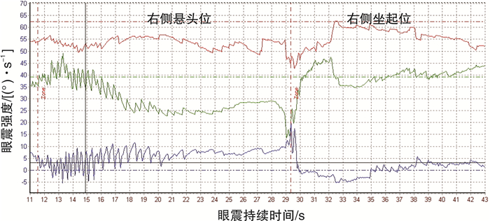
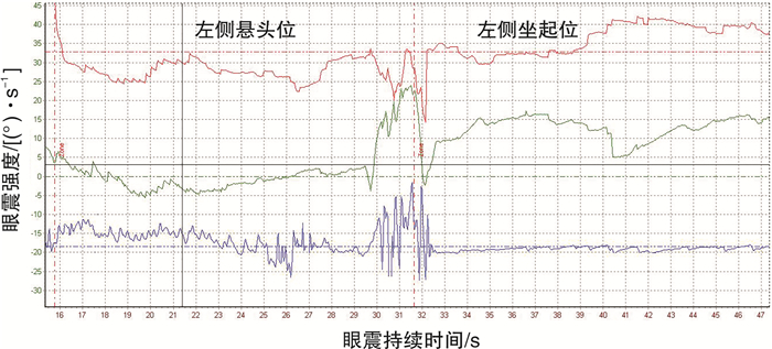
摘要: 目的 应用三维视频眼震图(3D-VNG),记录分析后半规管管石症(PSC-Can)诱发扭转眼震的三维方向特征。方法 回顾60例(左侧22例,右侧38例)PSC-Can患者在Dix-Hallpike试验患侧悬头位与坐起位诱发的垂直扭转眼震,分析其水平分量、垂直分量和扭转分量的眼震特征。结果 60例PSC-Can患者在Dix-Hallpike试验的患侧悬头位和坐起位均诱发出垂直扭转性眼震,在3D-VNG上均呈现水平、垂直、扭转三个眼震分量。在患侧悬头位的3D-VNG上,左/右PSC-Can眼震的水平分量46例向对侧(另14例向同侧)、垂直分量均向上、扭转分量则分别向上/向下;坐起位三个分量诱发眼震的强度弱于悬头位,且垂直和扭转分量眼震方向较悬头位均反转,水平分量眼震反转39例,不反转21例。结论 3D-VNG技术可以从水平、垂直、扭转三个眼震分量客观记录PSC-Can扭转特征性眼震,为PSC-Can诱发眼震的量化分析及半规管生理性功能研究提供了更客观、全面的信息。Abstract: Objective The three-dimensional direction feature of torsional nystagmus induced by posterior semicircular canal canalithasis (PSC-Can) was recorded and analyzed using three-dimensional video nystagmography (3D-VNG).Methods Sixty patients (22 on the left side and 38 on the right side) with PSC-Can were enrolled for torsional nystagmus evoked by Dix-Hallpike test in the affected-side head-hanging and sitting positions, and the direction characteristics of the horizontal, vertical and torsional components were analyzed.Results Vertical torsional nystagmus was induced in 60 PSC-Can patients in the head-hanging and sitting positions evoked by Dix-Hallpike test, respectively. Horizontal, vertical, and torsional components of were presented in the 3D-VNG. In the head-hanging position, the direction of horizontal component in the left/right PSC-Can nystagmus was contralateral in 46 cases(the other 14 cases were ipsilateral), the vertical component was upward, and the torsional component was upward/downward, respectively. The intensity of nystagmus induced in the three components in the sitting position is weaker than in the head-hanging position, and the direction of nystagmus was reversed in both vertical and torsional components compared with the head-hanging position. However, the direction of the horizontal component was reversed in 39 cases and not reversed in 21 cases in the sitting position.Conclusion The horizontal, vertical and torsional components of the torsional nystagmus in PSC-Can patients recorded by 3D-VNG, which provided more comprehensive and objective information for the analysis of PSC-Can and the study of semicircular canal physiological function.
-

-
表 1 PSC-Can患者Dix-Hallpike试验分别在3D-VNG和视窗观测的眼震特征
Dix-Hallpike试验 3D-VNG眼震方向 视窗观测眼震方向 受累半规管 悬头位 坐起位 悬头位 坐起位 垂直 水平 扭转 垂直 水平 扭转 扭转方向 垂直 扭转方向 垂直 右侧 上 左a) 下 下 右a) 上 逆时针 向上 顺时针 向下 R-PSC-Can 左侧 上 右a) 上 下 左a) 下 顺时针 向上 逆时针 向下 L-PSC-Can 注:a)PSC-Can患者Dix-Hallpike试验显示悬头位水平分量成分多指向健侧,坐起位多指向患侧。 -
[1] von Brevern M, Radtke A, Lezius F, et al. Epidemiology of benign paroxysmal positional vertigo: a population based study[J]. J Neurol Neurosurg Psychiatry, 2007, 78(7): 710-715.
[2] 中华耳鼻咽喉头颈外科杂志编辑委员会, 中华医学会耳鼻咽喉头颈外科学分会. 良性阵发性位置性眩晕诊断和治疗指南(2017)[J]. 中华耳鼻咽喉头颈外科杂志, 2017, 52(3): 173-177. https://cpfd.cnki.com.cn/Article/CPFDTOTAL-ZGZP201709006014.htm
[3] von Brevern M, Bertholon P, Brandt T, et al. Benign paroxysmal positional vertigo: Diagnostic criteria[J]. J Vestib Res, 2015, 25(3/4): 105-117.
[4] Bhattacharyya N, Gubbels SP, Schwartz SR, et al. Clinical Practice Guideline: Benign Paroxysmal Positional Vertigo(Update)[J]., Otolaryngol Head Neck Surg, 2017, 156(3_suppl): S1-S47. doi: 10.1177/0194599816689667
[5] 韦一, 刘湘, 曾莉. 对比视频眼震电图与裸眼检查对良性阵发性位置性眩晕的诊断价值[J]. 现代医学与健康研究电子杂志, 2022, 6(8): 95-98. https://www.cnki.com.cn/Article/CJFDTOTAL-XYJD202208029.htm
[6] 张波, 孙敬武. 良性阵发性位置性眩晕患者裸眼及视频眼震图下眼震特征及定位诊断分析[J]. 听力学及言语疾病杂志, 2012, 20(3): 235-237. doi: 10.3969/j.issn.1006-7299.2012.03.012
[7] 惠晶, 訾定京, 范秀博. 视频眼震电图在良性阵发性位置性眩晕患者诊断中的临床应用价值研究[J]. 陕西医学杂志, 2019, 48(5): 636-638, 643. doi: 10.3969/j.issn.1000-7377.2019.05.026
[8] Wagner J, Lehnen N, Glasauer S, et al. Prognosis of idiopathic downbeat nystagmus[J]. Ann N Y Acad Sci, 2009, 1164: 479-481. doi: 10.1111/j.1749-6632.2009.03767.x
[9] Cremer PD, Migliaccio AA, Pohl DV, et al. Posterior semicircular canal nystagmus is conjugate and its axis is parallel to that of the canal[J]. Neurology, 2000, 54(10): 2016-2020. doi: 10.1212/WNL.54.10.2016
[10] Morrow MJ, Sharpe JA. Torsional nystagmus in the lateral medullary syndrome[J]. Ann Neurol, 1988, 24(3): 390-398. doi: 10.1002/ana.410240307
[11] Wang ZI, Dell'Osso LF. Eye-movement-based assessment of visual function in patients with infantile nystagmus syndrome[J]. Optom Vis Sci, 2009, 86(8): 988-995. doi: 10.1097/OPX.0b013e3181b2f2ee
[12] 陈飞云, 陈太生, 温超, 等. 水平半规管良性阵发性位置性眩晕患者眼震参数客观特征[J]. 中华耳鼻咽喉头颈外科杂志, 2013, 48(8): 622-627. doi: 10.3760/cma.j.issn.1673-0860.2013.08.003
[13] Toshiaki Y, Yoshio O, Kayo S, et al. 3D analysis of nystagmus in peripheral vertigo[J]. Acta Otolaryngol, 1997, 117(2): 135-138. doi: 10.3109/00016489709117754
[14] Imai T, Takeda N, Ito M, et al. 3D analysis of benign positional nystagmus due to cupulolithiasis in posterior semicircular canal[J]. Acta Otolaryngol, 2009, 129(10): 1044-1049. doi: 10.1080/00016480802566303
[15] Yagi T, Koizumi Y. 3D analysis of cough-induced nystagmus in a patient with superior semicircular canal dehiscence[J]. Auris Nasus Larynx, 2009, 36(5): 590-593. doi: 10.1016/j.anl.2009.01.012
[16] Yagi T, Koizumi Y, Sugizaki K. 3D analysis of spontaneous nystagmus in early stage of vestibular neuritis[J]. Auris Nasus Larynx, 2010, 37(2): 167-172. doi: 10.1016/j.anl.2009.05.008
[17] 区永康, 郑亿庆, 陈玲, 等. 不同类型良性阵发性位置性眩晕的诊断和治疗[J]. 中华耳科学杂志, 2006, 4(4): 279-282. doi: 10.3969/j.issn.1672-2922.2006.04.010
[18] 王娜, 陈太生, 林鹏, 等. 良性阵发性位置性眩晕的眼震图研究[J]. 临床耳鼻咽喉头颈外科杂志, 2009, 23(13): 597-600. https://www.cnki.com.cn/Article/CJFDTOTAL-LCEH200913011.htm
[19] 徐开旭, 陈太生, 王巍等. 后半规管良性阵发性位置性眩晕患者眼震参数客观特征[J]. 中华耳鼻咽喉头颈外科杂志, 2019, 54(10): 729-733. doi: 10.3760/cma.j.issn.1673-0860.2019.10.005
[20] 崔湘凝, 冯永, 梅凌云, 等. 后半规管良性阵发性位置性眩晕患者在体位诱发试验中的眼震特征分析[J]. 临床耳鼻咽喉头颈外科杂志, 2015, 29(1): 27-30. https://www.cnki.com.cn/Article/CJFDTOTAL-LCEH201501010.htm
-


 下载:
下载:
