
Endoscopic management and outcome of nasosinusitis in non-traumatic dehiscence of the lamina papyracea with orbital content herniation
-
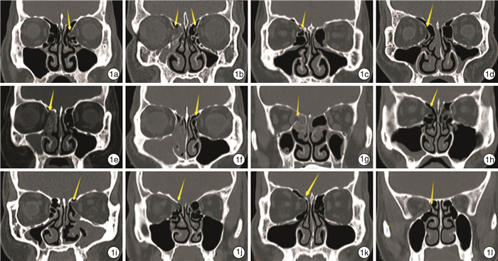
摘要: 目的 分析非创伤性纸样板缺损伴眶内容物疝出的鼻窦炎的CT表现、临床特征、内镜处理及结局。方法 回顾性分析福建医科大学附属第一医院耳鼻咽喉头颈外科2019年1月—2020年10月收治的诊断为慢性鼻窦炎或鼻中隔偏曲患者的临床资料,所有患者均排除既往颌面部或眼眶外伤史和鼻眼相关手术史,共纳入研究患者686例(男448例,女238例)。结果 12例患者被确诊为纸样板缺损,所有患者术前CT均显示纸样板缺损,缺损位置均局限于筛窦。随访期间未见复发。结论 对于合并纸样板缺损的鼻窦炎患者采取功能性鼻窦内镜手术,在精确熟练的手术操作、良好的出血控制和清晰的视野下可以将所有筛窦气房充分开放,术中确保未损伤疝出的眶周脂肪,术后给予适当的术腔填塞,可以避免眼眶相关并发症的发生,同时鼻窦炎也能得到很好的改善。Abstract: Objective To analyze the CT manifestations, clinical features, and endoscopic management and outcome of nasosinusitis in non-traumatic dehiscence of the lamina papyracea with herniation of orbital contents.Methods From January 2019 to October 2020, a total of 686 cases with chronic nasosinusitis or nasal septum deviation were admitted to our department, including 448 male cases and 238 female cases. No patient had prior maxillofacial or orbital trauma as well as surgery related to nose and eyes. The clinical data were retrospectively analyzed.Results Twelve patients were diagnosed as dehiscence of the lamina papyracea. Preoperative CT revealed that the location of dehiscence was only in the ethmoid sinus.Conclusion For nasosinusitis patients with non-traumatic dehiscence of the lamina papyracea, all ethmoid cells should be opened during FESS. Precise and skillful operation, good bleeding control and clear visual field were critical. no damage to the herniated periorbital fat during the operation and appropriate cavity packing after the operation are essential, which could avoid the orbital-related complications as well as improve the symptom resolution. No recurrence was found during the follow-up period.
-
Key words:
- dehiscence of the lamina papyracea /
- nasosinusitis /
- endoscopic
-

-
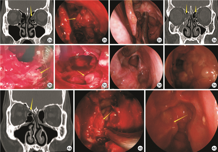
图 2 例1患者临床资料 2a:术前CT检查示左侧纸样板缺损;2b:术中内镜下在相应位置触及疝到筛窦腔的眶内容物;2c:术后3个月内镜复查,原先疝出位置组织变致密,未再触及明显薄弱位置,眶周结构完整;图3 例2患者临床资料 3a:术前CT影像示双侧纸样板缺损;3b、3c:术中内镜下在相应位置触及疝到筛窦腔的眶内容物;3d、3e:术后11个月内镜复查,原先疝出位置组织变致密,未再触及明显薄弱位置,眶周结构完整;图4 例3患者临床资料 4a:术前CT影像示右侧纸样板缺损;4b:术中内镜下在相应位置触及疝到筛窦腔的眶内容物;4c:术后9个月内镜复查,原先疝出位置组织变致密,未再触及明显薄弱位置,眶周结构完整。
表 2 12例纸样板缺损患者的临床资料
患者 性别 年龄 缺损位置 缺损程度 组别 1 男 55 左侧 Ⅰ级 慢性鼻窦炎 2 男 64 双侧 Ⅰ级 慢性鼻窦炎 3 男 36 右侧 Ⅰ级 慢性鼻窦炎 4 男 73 右侧 Ⅰ级 慢性鼻窦炎 5 女 57 右侧 Ⅰ级 慢性鼻窦炎 6 男 46 左侧 Ⅰ级 慢性鼻窦炎 7 男 55 右侧 Ⅰ级 慢性鼻窦炎 8 男 66 右侧 Ⅱ级 慢性鼻窦炎 9 女 46 左侧 Ⅰ级 慢性鼻窦炎 10 男 41 右侧 Ⅰ级 鼻中隔偏曲 11 男 57 右侧 Ⅲ级 鼻中隔偏曲 12 女 43 右侧 Ⅰ级 鼻中隔偏曲  下载: 导出CSV
-
[1] Kitaguchi Y, Takahashi Y, Mupas-Uy J, et al. Characteristics of Dehiscence of Lamina Papyracea Found on Computed Tomography Before Orbital and Endoscopic Endonasal Surgeries[J]. J Craniofac Surg, 2016, 27(7): e662-e665. doi: 10.1097/SCS.0000000000003005
[2] Han MH, Chang KH, Min YG, et al. Nontraumatic prolapse of the orbital contents into the ethmoid sinus: evaluation with screening sinus CT[J]. Am J Otolaryngol, 1996, 17(3): 184-189. doi: 10.1016/S0196-0709(96)90058-7
[3] Seeley MJ, Waterhouse DR, Shetty S, et al. Boundary issues: a case of nontraumatic bilateral dehiscence of the lamina papyracea[J]. Arch Otolaryngol Head Neck Surg, 2010, 136(1): 88-89. doi: 10.1001/archoto.2009.196
[4] Gerard M, Merle H, Domenjôd M, ,et al. Isolated blow out fracture of the medial wall of the orbit with medial rectus entrapment. Apropos of 3 cases[J]. J Fr Ophtalmol, 1996, 19(10): 591-596.
[5] 王雪峰, 陈冬, 李兵, 等. 内镜下修复眶内容击出性骨折伴损伤性视神经病变1例[J]. 临床耳鼻咽喉头颈外科杂志, 2009, 23(14): 664-664. https://www.cnki.com.cn/Article/CJFDTOTAL-LCEH200914020.htm
[6] Meyers RM, Valvassori G. Interpretation of anatomic variations of computed tomography scans of the sinuses: a surgeon's perspective[J]. Laryngoscope, 1998, 108(3): 422-425. doi: 10.1097/00005537-199803000-00020
[7] Moulin G, Dessi P, Chagnaud C, et al. Dehiscence of the lamina papyracea of the ethmoid bone: CT findings[J]. AJNR Am J Neuroradiol, 1994, 15(1): 151-153.
[8] René C. Update on orbital anatomy[J]. Eye(Lond), 2006, 20(10): 1119-1129.
[9] Shpilberg KA, Daniel SC, Doshi AH, et al. CT of Anatomic Variants of the Paranasal Sinuses and Nasal Cavity: Poor Correlation With Radiologically Significant Rhinosinusitis but Importance in Surgical Planning[J]. AJR Am J Roentgenol, 2015, 204(6): 1255-1260. doi: 10.2214/AJR.14.13762
[10] Papadopoulou AM, Chrysikos D, Samolis A, et al. Anatomical Variations of the Nasal Cavities and Paranasal Sinuses: A Systematic Review[J]. Cureus, 2021, 13(1): e12727.
[11] Yang YX, Lu QK, Liao JC, et al. Morphological characteristics of the anterior ethmoidal artery in ethmoid roof and endoscopic localization[J]. Skull Base, 2009, 19(5): 311-317. doi: 10.1055/s-0028-1115323
[12] Chao TK. Protrusion of orbital content through dehiscence of lamina papyracea mimics ethmoiditis: a case report[J]. Otolaryngol Head Neck Surg, 2003, 128(3): 433-435. doi: 10.1067/mhn.2003.105
[13] Chao TK. Uncommon anatomic variations in patients with chronic paranasal sinusitis[J]. Otolaryngol Head Neck Surg, 2005, 132(2): 221-225. doi: 10.1016/j.otohns.2004.09.132
-
图(2)
表(1)
计量
- 文章访问数: 683
- PDF下载数: 171
- 施引文献: 0