The value of ACR-TIRADS and C-TIRADS in the diagnosis of nodular Hashimoto thyroiditis and papillary thyroid carcinoma with Hashimoto thyroiditis
-
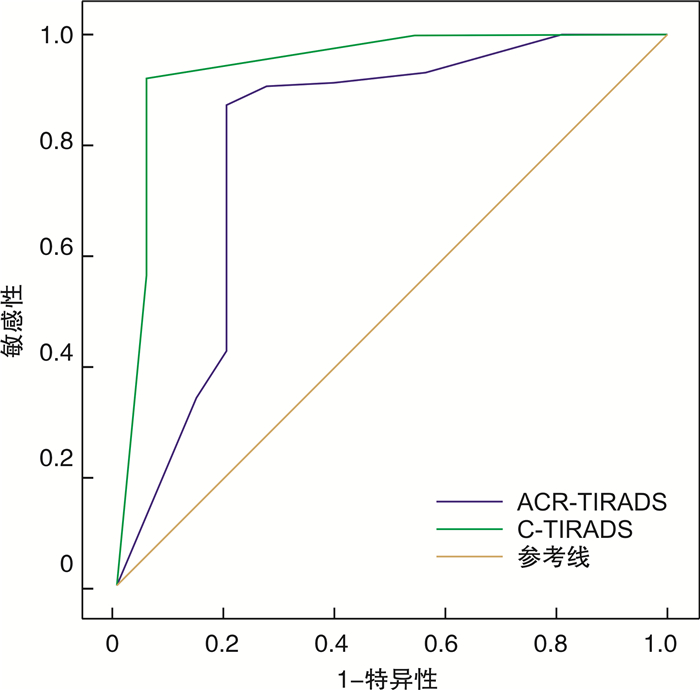
摘要: 目的 探讨美国放射学会甲状腺影像报告与数据系统(ACR-TIRADS)与中国甲状腺结节超声恶性危险分层(C-TIRADS)对桥本甲状腺炎背景下桥本结节和甲状腺乳头状癌的诊断价值。方法 回顾性分析2018年8月-2021年5月于河北北方学院附属第一医院行甲状腺超声检查并经手术病理证实的桥本甲状腺炎伴有结节或甲状腺乳头状癌患者141例(204个结节),对所有结节行超声检查,并且按照ACR-TIRADS和C-TIRADS的分类标准对204个结节进行评分分级,以手术病理结果 为金标准,构建ACR-TIRADS和C-TIRADS评估桥本结节和甲状腺乳头状癌性质的受试者工作特征曲线,分析和比较二者的诊断效能。结果 ① 超声特征结果显示桥本结节和甲状腺乳头状癌在分布位置、回声、钙化和边缘之间的差异有统计学意义(P < 0.001),二者在结构和纵横比上差异无统计学意义(P=0.141,P=0.240),桥本结节多表现为无局灶性强回声和高/等回声,甲状腺乳头状癌多表现为局灶性强回声和甲状腺外侵犯; ②C-TIRADS诊断桥本结节和甲状腺乳头状癌性质的敏感性和阴性预测值分别为91.7%和83.1%,均高于ACR-TIRADS,差异存在统计学意义(P=0.021,P=0.013);特异性和阳性预测值分别为98.3%和99.2%,均略高于ACR-TIRADS,差异无统计学意义(P=0.157,P=0.062)。ACR-TIRADS和C-TIRADS两种超声指南的曲线下面积分别为0.806和0.941,差异有统计学意义(P=0.031);③C-TIRADS超声指南不必要细针抽吸活检率为10.3%,低于ACR-TIRADS。结论 C-TIRADS对桥本甲状腺炎背景下的桥本结节和甲状腺乳头状癌的诊断具有较高的价值,有助于临床对此类结节的评估。Abstract: Objective To explore the diagnostic value of American Society of Radiology Thyroid Imaging Reporting and Data System(ACR-TIRADS) and Chinese Thyroid Nodule Ultrasound Malignant Risk Stratification(C-TIRADS) in nodular Hashimoto thyroiditis and papillary thyroid carcinoma with Hashimoto thyroiditis.Methods This retrospective analysis included 144 patients(204 thyroid nodules) accompanied by nodular Hashimoto thyroiditis or papillary thyroid carcinoma under the background of Hashimoto thyroiditis confirmed by surgical pathology examination in the First Affiliated Hospital of Hebei North University from August 2018 to May 2021, all nodules were examined by ultrasound, and 204 nodules were scored and graded according to the classification standards of ACR-TIRADS and C-TIRADS. The surgical pathological results were the gold standard. The receiver operating characteristic curve of ACR-TIRADS and C-TIRADS was constructed to evaluate and compare the diagnostic performance of the two guideline.Results ① Ultrasound feature results showed that nodular Hashimoto thyroiditis and Papillary thyroid carcinoma had statistically significant differences in the location, echogenicity, calcifications and margins(P < 0.001), but there is no significant difference in structure and aspect ratio between the two kinds of nodular(P=0.141, P=0.240); nodular Hashimoto thyroiditis were mostly absent focal echogenicity and hyperechogenicity, while papillary thyroid carcinoma was mostly manifested as focal echogenicity and extrinsic thyroid invasion. ②The sensitivity and negative predictive value of C-TIRADS were 91.7% and 83.1%, respectively, which were higher than those of ACR-TIRADS, and the difference was statistically significant(P=0.021, P=0.013); The specificity and positive predictive value of C-TIRADS T were 98.3% and 99.2%, both of which were slightly higher than ACR-TIRADS, althought the difference was not statistically significant(P=0.157, P=0.062). The area under the curve of the ACR-TIRADS and C-TIRADS were 0.806 and 0.941, respectively, and the difference was statistically significant(P=0.031). ③The unnecessary FNAB rate of C-TIRADS was 10.3%, which was lower than ACR-TIRADS.Conclusion C-TI-RADS has a better diagnostic value of nodular Hashimoto thyroiditis and thyroid papillary carcinoma under the background of Hashimoto thyroiditis, which is helpful for clinical evaluation of such nodules.
-

-
表 1 ACR-TIRADS和C-TIRADS对甲状腺结节行细针抽吸活检标准[7]
指南 行细针抽吸活检标准 ACR-TIRADS 良性 否 非可疑恶性 否 低度可疑恶性 推荐结节尺寸≥25 mm; 观察≥15 mm 中度可疑恶性 推荐结节尺寸≥15 mm; 观察≥10 mm 高度可疑恶性 推荐结节尺寸≥10 mm; 观察≥5 mm C-TIRADS 良性 否 良性可能 否 低度可疑恶性 推荐结节尺寸≥15 mm; 推荐≥10 mm(存在一个可疑恶性超声特征) 中度可疑恶性 推荐结节尺寸≥10 mm; 推荐≥5 mm(存在一个可疑恶性超声特征) 高度可疑恶性 推荐结节尺寸≥10 mm; 推荐≥5 mm(存在一个可疑恶性超声特征) 高度提示恶性 推荐结节尺寸≥10 mm; 推荐≥5 mm(存在一个可疑恶性超声特征); 推荐(存在二个可疑恶性超声特征) 表 2 桥本甲状腺炎背景下桥本结节和甲状腺乳头状癌的超声特征比较
项目 数量 桥本结节 甲状腺乳头状癌 P值 项目 数量 桥本结节 甲状腺乳头状癌 P值 例数 141 52 89 回声 204 60 144 < 0.001 平均年龄/岁 47.1± 49.7± 48.7± >0.05 高/等回声 40 36 4 8.2 12.2 11.2 低回声 118 16 102 性别 极低回声 44 6 38 男 11 0 11 < 0.05 无回声 2 2 0 女 130 52 78 >0.05 边缘 204 60 144 < 0.001 结节/个 204 60 144 光整 14 14 0 平均直径/mm 17.9± 15.1± 21.2± >0.05 不规则 36 15 21 10.1 10.6 7.6 模糊 95 30 65 分布位置 204 60 144 < 0.001 甲状腺外侵犯 59 1 58 上部 30 8 22 钙化 204 60 144 < 0.001 中部 46 14 32 微钙化 92 2 90 下部 40 30 10 彗星尾伪像 8 8 0 峡部 88 8 80 粗钙化 29 2 27 结构 204 60 144 0.141 周边钙化 13 0 13 实性 191 56 135 无局灶性强回声 69 48 21 囊实性部分 11 2 9 纵横比 204 60 144 0.240 囊性 1 1 0 ≥1 63 15 48 海绵状 1 1 0 < 1 141 45 96 表 3 ACR-TIRADS和C-TIRADS诊断桥本甲状腺炎背景下结节性质的情况比较
超声指南 桥本结节 甲状腺乳头状癌 结节数量 建议恶性率 计算恶性率 P值 ACR-TIRADS 60 144 204 P < 0.001 良性 5 0 5 ≤2 0 非可疑恶性 6 0 6 ≤2 0 低度可疑恶性 23 1 24 < 5 4.2 中度可疑恶性 26 3 29 5~20 10.3 高度可疑恶性 1 140 141 >20 99.3 C-TIRADS 60 144 204 P < 0.001 良性 1 0 1 0 0 良性可能 2 0 2 < 2 0 低度可疑恶性 21 2 23 2~10 8.7 中度可疑恶性 30 10 40 10~50 25 高度可疑恶性 5 41 46 50~90 89.1 高度提示恶性 1 91 92 >90 98.9 表 4 ACR-TIRADS和C-TIRADS对桥本甲状腺炎背景下桥本结节和甲状腺乳头状癌的诊断效能
超声指南 诊断截点/分 敏感性/% 特异性/% 准确性/% 阳性预测值/% 阴性预测值/% ACR-TIRADS 6.5 86.8(125/144) 95.0(57/60) 89.2(182/204) 97.7(125/128) 75.0(57/76) C-TIRADS 3.0 91.7(132/144) 98.3(59/60) 93.6(191/204) 99.2(132/133) 83.1(59/71) 表 5 ACR-TIRADS和C-TIRADS不必要细针抽吸活检率的比较
超声指南 细针抽吸活检 良性结节中行细针抽吸活检/% 恶性结节中行细针抽吸活检/% 不必要细针抽吸活检率/% 细针抽吸活检假阳性率/% ACR-TIRADS 132 15.9(21/132) 84.1(111/132) 10.3(21/204) 35(21/60) C-TIRADS 144 10.4(15/144) 89.6(129/144) 7.4(15/204) 25(15/60) -
[1] 林婉玲, 雷志锴, 丁金旺, 等. 预测桥本甲状腺炎背景下甲状腺乳头状癌超声特征与中央区淋巴结转移的价值[J]. 浙江医学, 2021, 43(7): 748-752. https://www.cnki.com.cn/Article/CJFDTOTAL-ZJYE202107014.htm
[2] 吴翠怡, 冀将婷, 周美君, 等. ACR TI-RADS诊断桥本甲状腺炎背景下桥本结节与甲状腺乳头状癌的价值[J]. 中国临床医学影像杂志, 2021, 32(4): 245-249. https://www.cnki.com.cn/Article/CJFDTOTAL-LYYX202104008.htm
[3] 杨涛, 朱世琴, 刘慧, 等. VTIQ技术对桥本甲状腺炎合并乳头状癌颈部中央区淋巴结性质判定的价值[J]. 中国超声医学杂志, 2021, 37(7): 729-732. doi: 10.3969/j.issn.1002-0101.2021.07.003
[4] 李潜, 丁思悦, 郭兰伟, 等. 甲状腺结节超声恶性危险分层中国指南(C-TIRADS)联合人工智能辅助诊断对甲状腺结节鉴别诊断的效能评估[J]. 中华超声影像学杂志, 2021, 30(3): 231-235. doi: 10.3760/cma.j.cn131148-20201106-00858
[5] 周建桥, 詹维伟. 2020年中国超声甲状腺影像报告和数据系统(C-TIRADS)指南解读[J]. 诊断学理论与实践, 2020, 19(4): 350-353. https://www.cnki.com.cn/Article/CJFDTOTAL-ZDLS202004008.htm
[6] Zhu H, Yang Y, Wu S, et al. Diagnostic performance of US-based FNAB criteria of the 2020 Chinese guideline for malignant thyroid nodules: comparison with the 2017 American College of Radiology guideline, the 2015 American Thyroid Association guideline, and the 2016 Korean Thyroid Association guideline[J]. Quant Imaging Med Surg, 2021, 11(8): 3604-3618. doi: 10.21037/qims-20-1365
[7] Tessler FN, Middleton WD, Grant EG, et al. ACR Thyroid Imaging, Reporting and Data System(TI-RADS): White Paper of the ACR TI-RADS Committee[J]. J Am Coll Radiol, 2017, 14(5): 587-595. doi: 10.1016/j.jacr.2017.01.046
[8] 胡梅, 李明星, 王世界, 等. 伴桥本甲状腺炎的甲状腺良恶性结节: 超声特征及甲状腺超声征象报告与数据系统诊断价值[J]. 中国医学影像技术, 2019, 35(6): 828-832. https://www.cnki.com.cn/Article/CJFDTOTAL-ZYXX201906011.htm
[9] Remonti LR, Kramer CK, Leitão CB, et al. Thyroid ultrasound features and risk of carcinoma: a systematic review and meta-analysis of observational studies[J]. Thyroid, 2015, 25(5): 538-550. doi: 10.1089/thy.2014.0353
[10] 侯佳欣, 李茂萍, 彭晓琼, 等. 桥本甲状腺炎对≥1 cm甲状腺结节超声引导下细针穿刺细胞学检查诊断效能的影响[J]. 临床耳鼻咽喉头颈外科杂志, 2021, 35(9): 807-812. https://www.cnki.com.cn/Article/CJFDTOTAL-LCEH202109008.htm
[11] Wu H, Zhang B. Ultrasonographic appearance of focal Hashimoto's thyroiditis: A single institution experience[J]. Endocr J, 2015, 62(7): 655-663. doi: 10.1507/endocrj.EJ15-0083
[12] 薛恒, 陈文, 沈伟伟, 等. 甲状腺影像报告与数据系统(TI-RADS)观察者一致性与阳性预测值的研究[J]. 中华超声影像学杂志, 2018, 27(5): 401-405. doi: 10.3760/cma.j.issn.1004-4477.2018.05.007
[13] Wu H, Zhang B, Li J, et al. Echogenic foci with comet-tail artifact in resected thyroid nodules: Not an absolute predictor of benign disease[J]. PLoS One, 2018, 13(1): e0191505. doi: 10.1371/journal.pone.0191505
-


 下载:
下载: