
The value of magnetic resonance hydrography in the localization of fistula in patients with cerebrospinal fluid rhinorrhea
-
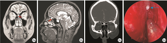
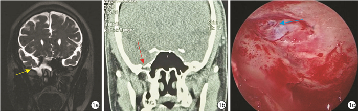
摘要: 目的 通过比较脑脊液鼻漏患者术前核磁共振水成像(MRH)、鼻窦冠状位CT判断的漏口位置与术中内镜所见漏口位置的一致性,评价MRH在术前漏口定位过程中的应用价值。方法 回顾性分析2015年2月-2021年2月天津市环湖医院收治的脑脊液鼻漏患者58例,采用鼻窦冠状位CT、MRH及MRH结合鼻窦冠状位CT这三种方法分别判断漏口位置,比较不同方法确定的漏口位置与术中漏口位置的一致性。结果 MRH结合鼻窦冠状位CT的诊断阳性率(91.4%)明显高于MRH阳性率(74.1%),差异有统计学意义(P < 0.05)。MRH诊断阳性率(74.1%)高于常规鼻窦冠状位CT检查阳性率(43.1%),差异有统计学意义(P < 0.01)。结论 影像学检查技术的综合运用对脑脊液鼻漏的正确诊断有着重要的临床指导意义,术前准确定位才能更有针对性地进行精确的修补手术治疗。MRH结合鼻窦冠状位CT可以明显提高漏口位置的诊断率,因此可以作为脑脊液鼻漏患者术前定位的首选方法。Abstract: Objective To evaluate the application value of magnetic resonance hydrography in determine the leak location of patients with cerebrospinal fluid rhinorrhea.Methods Clinical data of 58 patients with cerebrospinal fluid rhinorrhea treated in Tianjin Huanhu Hospital from February 2015 to February 2021 were analyzed retrospectively. The leak position was judged by three methods: nasal sinus coronal CT, magnetic resonance hydrography and magnetic resonance hydrography combined with nasal sinus coronal CT. The consistency of the leak position among groups determined by different imageological examination pre-operation was compared with the real leak position found during the operation.Results The positive rate of magnetic resonance hydrography combined with coronal CT in paranasal sinuses(91.4%) was significantly higher than that of magnetic resonance hydrography(74.1%)(P>0.05). The positive rate of magnetic resonance hydrography(74.1%) was higher than that of CT(43.1%)(P>0.01).Conclusion The comprehensive application of imaging examination technology has important clinical guiding significance for the correct diagnosis of cerebrospinal fluid rhinorrhea. Magnetic resonance hydrography combined with coronal CT of paranasal sinus can effectively find the leak location, which was suggested as the first choice for preoperative localization of cerebrospinal fluid rhinorrhea.
-

-
表 1 不同位置脑脊液鼻漏不同方法的阳性例数和诊断率
例(%) 位置 MRH阳性 冠状位CT阳性 MRH结合冠状位CT阳性 筛窦 18(60.0) 8(26.7) 27(90.0) 额窦 5(83.3) 3(50.0) 5(83.3) 蝶窦外侧隐窝 13(100.0) 8(61.5) 13(100.0) 蝶窦鞍底 7(77.8) 6(66.7) 8(88.2)  下载: 导出CSV
-
[1] Caballero N, Bhalla V, Stankiewicz JA, et al. Effect of lumbar drain placement on recurrence of cerebrospinal rhinorrhea after endoscopic repair[J]. Int Forum Allergy Rhinol, 2012, 2(3): 222-226. doi: 10.1002/alr.21023
[2] Majhi S, Sharma A. Outcome of Endoscopic Cerebrospinal Fluid Rhinorrhoea Repair: An Institutional Study[J]. Indian J Otolaryngol Head Neck Surg, 2019, 71(1): 76-80. doi: 10.1007/s12070-018-1485-2
[3] Daudia A, Biswas D, Jones NS. Risk of meningitis with cerebrospinal fluid rhinorrhea[J]. Ann Otol Rhinol Laryngol, 2007, 116(12): 902-905. doi: 10.1177/000348940711601206
[4] Miner JR, Heegaard W, Mapes A, et al. Presentation, time to antibiotics, and mortality of patients with bacterial meningitis at an urban county medical center[J]. J Emerg Med, 2001, 21(4): 387-392. doi: 10.1016/S0736-4679(01)00407-3
[5] Ozturk O, Polat S, Uneri C. Endoscopic endonasal management of cerebrospinal fluid rhinorrhea[J]. J Craniofac Surg, 2012, 23(4): 1087-1092. doi: 10.1097/SCS.0b013e31824e6a44
[6] 朱正洁, 程岚, 杨军. 自发性脑脊液鼻漏的发病机制及治疗[J]. 临床耳鼻咽喉头颈外科杂志, 2020, 34(6): 573-576. https://www.cnki.com.cn/Article/CJFDTOTAL-LCEH202006023.htm
[7] Sivanandapanicker J, Nagar M, Kutty R, et al. Analysis and Clinical Importance of Skull Base Fractures in Adult Patients with Traumatic Brain Injury[J]. J Neurosci Rural Pract, 2018, 9(3): 370-375. doi: 10.4103/jnrp.jnrp_38_18
[8] Oakley GM, Alt JA, Schlosser RJ, et al. Diagnosis of cerebrospinal fluid rhinorrhea: an evidence-based review with recommendations[J]. Int Forum Allergy Rhinol, 2016, 6(1): 8-16. doi: 10.1002/alr.21637
[9] Hiremath SB, Gautam AA, Sasindran V, et al. Cerebrospinal fluid rhinorrhea and otorrhea: A multimodality imaging approach[J]. Diagn Interv Imaging, 2019, 100(1): 3-15. doi: 10.1016/j.diii.2018.05.003
[10] Reddy M, Baugnon K. Imaging of Cerebrospinal Fluid Rhinorrhea and Otorrhea[J]. Radiol Clin North Am, 2017, 55(1): 167-187. doi: 10.1016/j.rcl.2016.08.005
[11] Tuntiyatorn L, Laothammatas J. Evaluation of MR cisternography in diagnosis of cerebrospinal fluid fistula[J]. J Med Assoc Thai, 2004, 87(12): 1471-1416.
[12] Ecin G, Oner AY, Tokgoz N, et al. T2-weighted vs. intrathecal contrast-enhanced MR cisternography in the evaluation of CSF rhinorrhea[J]. Acta Radiol, 2013, 54(6): 698-701. doi: 10.1177/0284185113478008
[13] Payne RJ, Frenkiel S, Glikstein R, et al. Role of computed tomographic cisternography in the management of cerebrospinal fluid rhinorrhea[J]. J Otolaryngol, 2003, 32(2): 93-100. doi: 10.2310/7070.2003.37260
[14] 栾宁, 朱丽, 徐驰宇. 脑脊液鼻漏的定位及修补——附12例报告[J]. 中国微创外科杂志, 2019, 19(12): 1132-1135. doi: 10.3969/j.issn.1009-6604.2019.12.019
[15] 杨邦坤, 聂颖, 秦军, 等. 脑脊液鼻漏的诊断和治疗分析(附84例报告)[J]. 中国临床神经外科杂志, 2014, 4(19): 203-205. https://cdmd.cnki.com.cn/Article/CDMD-10487-1013012936.htm
[16] Liao KH, Wang JY, Lin HW, et al. Risk of death in patients with post-traumatic cerebrospinal fluid leakage-analysis of 1773 cases[J]. J Chin Med Assoc, 2016, 79(2): 58-64. doi: 10.1016/j.jcma.2015.10.002
[17] 张胜男, 姜彦, 于龙刚, 等. 鼻内镜下脑脊液鼻漏修补术诊疗分析[J]. 临床耳鼻咽喉头颈外科杂志, 2019, 33(12): 1189-1195, 1199. https://www.cnki.com.cn/Article/CJFDTOTAL-LCEH201912019.htm
[18] Shetty PG, Shroff MM, Fatterpekar GM, et al. A retrospective analysis of spontaneous sphenoid sinus fistula: MR and CT findings[J]. AJNR Am J Neuroradiol, 2000, 21(2): 337-342.
-
图(2)
表(1)
计量
- 文章访问数: 895
- PDF下载数: 475
- 施引文献: 0