Application of esophageal CT to establish the evaluation model of foreign body position in rigid esophagoscopic surgery
-
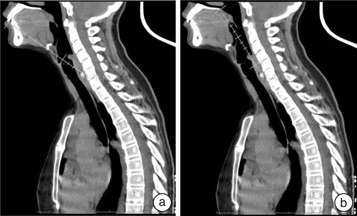
摘要: 目的 应用食管异物CT的相关径线建立与食管镜手术中异物真实位置的相关模型。方法 选取就诊于首都医科大学附属北京同仁医院耳鼻咽喉头颈外科急诊,经食管CT确诊为食管异物的患者33例。测量食管CT相关径线(气道长度、舌骨前缘-下颌骨距离、门齿延长线-后鼻嵴、异物距硬腭距离、异物距门齿距离、前后鼻嵴连线-脊柱线夹角、前后鼻嵴线与气道长度线夹角、下颌骨最低点-舌骨最高点-脊柱线夹角)、记录患者的身高、体重、BMI等,术中患者头部充分后仰,经口置入硬质食管镜,食管镜前端接触异物时记录置入食管镜距离门齿的距离。应用多元线性分析的方法建立食管CT相关径线与术中异物距门齿距离的模型。结果 食管异物最常见的异物为枣核(14例),其次为鱼刺(13例);食管CT测量的异物距硬腭、异物距门齿的距离均小于术中异物距门齿的实际距离(P < 0.001),差异有统计学意义;多元线性回归分析发现患者BMI(P=0.037)和异物距硬腭(P < 0.001)的距离与术中的异物距门齿的实际距离具有相关性。LR=3.708+0.130×BMI+0.857×Lct(cm),R2=0.736,调整后的R2=0.719。结论 通过食管CT测量异物距硬腭的距离结合患者BMI可以预测全身麻醉硬质食管镜手术中异物距门齿的真实距离,对手术中探查寻找异物具有一定参考价值。Abstract: Objective Establish a correlation model with the true position of the foreign body in the esophageal foreign body surgery using the relevant diameter of the esophageal foreign body computed tomography(CT).Methods Thirty-three patients who were diagnosed with esophageal foreign bodies by esophageal CT in the emergency department of the Department of Otolaryngology Head and Neck Surgery, Beijing Tongren Hospital, Capital Medical University, were selected to measure the CT-related diameters of the esophageal tube(airway length, hyoid anterior edge-mandibular distance, incisor extension line-Posterior nasal ridge, distance from foreign body to hard jaw, distance from foreign body to incisor, front and back nasal crest line-spine line included angle, front and back nasal crest line and airway length line included angle, the lowest point of mandible-highest point of hyoid bone-and Spine angle), record the height and weight of the patient and calculate the body mass index(BMI). During the operation, the patient's head is fully tilted back, and the rigid esophagus is inserted through the mouth, and the front end of the esophagus is recorded when it touches a foreign body. The method of multivariate linear analysis was used to calculate the CT diameter that correlated with the distance between the foreign body and the incisor during the operation.Results The most common foreign body in the esophagus is jujube pit(14 cases), followed by fish bones(13 cases); the distance between the foreign body and the hard jaw, the incisor teeth measured by CT of the esophagus is less than the actual distance between the foreign body and the incisor during the operation(P < 0.001), the difference was statistically significant. Multiple linear regression analysis found that the patient's BMI(P=0.037) and the distance of the foreign body from the hard jaw(P < 0.001) were correlated with the actual distance of the foreign body from the incisor during the operation. LR=3.708+0.130×BMI+0.857×Lct(cm), R2=0.736, adjusted R2=0.719.Conclusion The distance between the foreign body and the hard jaw measured by esophageal CT combined with the patient's BMI can predict the distance of the foreign body during rigid esophagoscopic surgery under general anesthesia and provide a certain reference value for the detection of foreign body during the operation.
-
Key words:
- esophageal foreign body /
- computed tomography /
- rigid esophagoscopy
-

-
表 1 食管异物的患者信息及种类、位置
患者 性别 年龄/岁 BMI 异物种类 距门齿/cm 距硬腭/cm 术中距门齿/cm 1 女 62 22.0 枣核 13.2 11.8 16.0 2 女 29 18.8 枣核 13.3 11.3 16.2 3 男 31 24.4 鸭骨头 17.3 17.3 21.8 4 男 63 32.9 鸡骨头 15.8 15.6 20.0 5 女 59 28.1 枣核 12.2 9.3 15.5 6 女 51 25.0 枣核 13.0 11.1 15.5 7 女 68 21.2 鱼骨 12.9 10.7 15.5 8 女 63 27.3 铝箔片 13.5 11.5 17.8 9 女 67 21.6 枣核 12.5 10.7 15.0 10 女 78 20.3 枣核 12.6 10.6 14.0 11 女 55 22.9 鱼刺 11.9 10.2 14.8 12 男 65 26.4 鱼刺 16.1 14.5 18.1 13 男 44 23.5 鱼骨 15.8 14.1 20.1 14 男 59 26.0 贝壳 17.3 16.6 24.5 15 女 31 18.2 鱼刺 12.6 9.8 14.3 16 男 25 32.4 鱼刺 14.3 11.9 16.9 17 男 53 17.3 鱼刺 14.5 12.8 15.5 18 女 64 25.0 鱼刺 13.2 11.1 20.0 19 男 44 26.1 枣核 15.8 14.8 19.2 20 女 56 26.4 鸡骨头 13.6 12.3 17.2 21 女 57 21.2 枣核 13.4 11.7 16.5 22 女 67 25.6 枣核 11.2 10.5 16.4 23 女 73 25.6 枣核 12.5 11.3 17.0 24 女 61 24.0 枣核 13.9 12.0 17.8 25 女 65 28.6 枣核 13.5 12.5 16.5 26 女 45 23.7 鱼刺 12.9 11.1 17.0 27 女 49 26.7 鱼刺 11.9 10.3 16.0 28 女 46 21.8 鱼刺 11.3 10.7 16.4 29 女 71 29.7 鱼刺 13.1 11.6 19.0 30 男 78 23.9 枣核 13.9 12.8 18.5 31 男 49 23.4 鸡骨头 16.2 16.3 19.5 32 女 57 17.2 鱼刺 11.4 10.6 15.5 33 女 69 20.7 枣核 10.0 9.3 14.0 -
[1] Lee CY, Kao BZ, Wu CS, et al. Retrospective analysis of endoscopic management of foreign bodies in the upper gastrointestinal tract of adults[J]. J Chin Med Assoc, 2019, 82(2): 105-109. doi: 10.1097/JCMA.0000000000000010
[2] Klein A, Ovnat-Tamir S, Marom T, et al. Fish Bone Foreign Body: The Role of Imaging[J]. Int Arch Otorhinolaryngol, 2019, 23(1): 110-115. doi: 10.1055/s-0038-1673631
[3] Xia Y, Zhang F, Xu H, et al. Use of the blue cotton screen method with endoscopy to detect occult esophageal foreign bodies[J]. Wideochir Inne Tech Maloinwazyjne, 2017, 12(4): 428-436.
[4] Hsieh A, Hsiehchen D, Layne S, et al. Trends and clinical features of intentional and accidental adult foreign body ingestions in the United States, 2000 to 2017[J]. Gastrointest Endosc, 2020, 91(2): 350-357 e1. doi: 10.1016/j.gie.2019.09.010
[5] Chirica M, Kelly MD, Siboni S, et al. Esophageal emergencies: WSES guidelines[J]. World J Emerg Surg, 2019, 14: 26. doi: 10.1186/s13017-019-0245-2
[6] Aiolfi A, Ferrari D, Riva CG, et al. Esophageal foreign bodies in adults: systematic review of the literature[J]. Scand J Gastroenterol, 2018, 53(10/11): 1171-1178.
[7] Shi WS, Su ZY, Wei CY, et al. Clinical features and standardized diagnosis and treatment of esophageal foreign bodies[J]. World Chin J Digestol, 2017, 25(30): 2721-2730. doi: 10.11569/wcjd.v25.i30.2721
[8] 王振晓, 曹晓明, 葛鑫颖, 等. 食管异物234例临床分析[J]. 临床耳鼻咽喉头颈外科杂志, 2019, 33(2): 148-151. https://www.cnki.com.cn/Article/CJFDTOTAL-LCEH201902013.htm
[9] Ruan WS, Li YN, Feng MX, et al. Retrospective observational analysis of esophageal foreign bodies: a novel characterization based on shape[J]. Sci Rep, 2020, 10(1): 4273. doi: 10.1038/s41598-020-61207-8
[10] Ferrari D, Aiolfi A, Bonitta G, et al. Flexible versus rigid endoscopy in the management of esophageal foreign body impaction: systematic review and meta-analysis[J]. World J Emerg Surg, 2018, 13: 42. doi: 10.1186/s13017-018-0203-4
[11] Adjeso T, Issaka A, Yabasin IB. Review of rigid esophagoscopy in a Tertiary Hospital in Ghana[J]. Pan Afr Med J, 2021, 39: 64.
[12] Yang W, Milad D, Wolter NE, et al. Systematic review of rigid and flexible esophagoscopy for pediatric esophageal foreign bodies[J]. Int J Pediatr Otorhinolaryngol, 2020, 139: 110397. doi: 10.1016/j.ijporl.2020.110397
-


 下载:
下载: