-
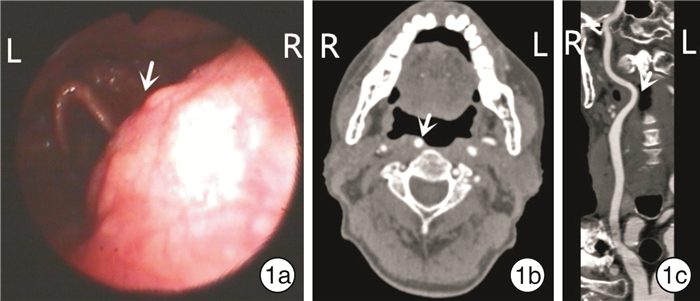
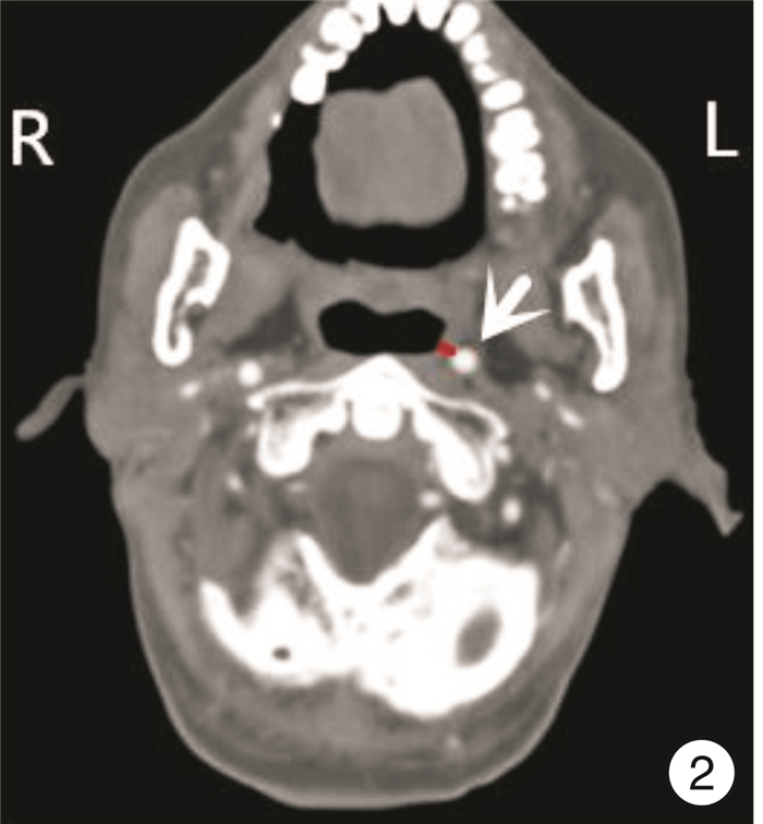
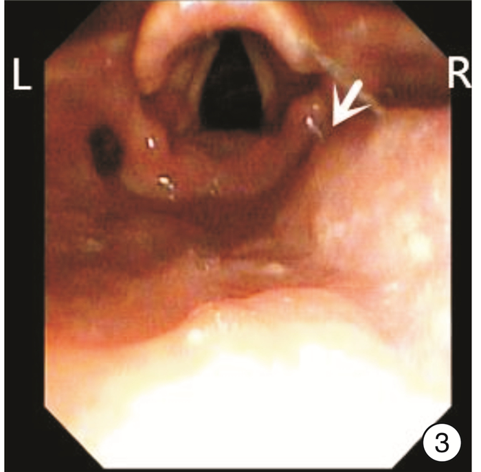
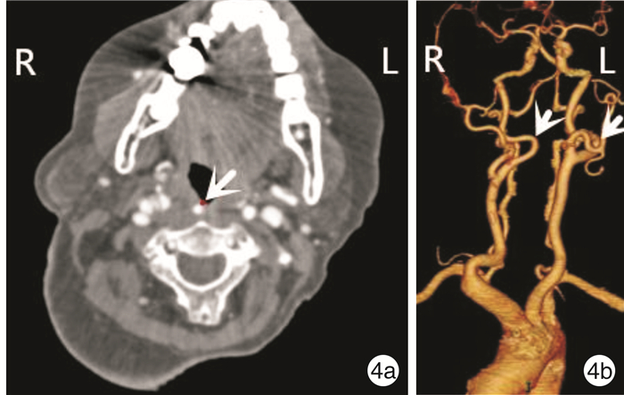
摘要: 目的 阐述颈段颈内动脉畸形的临床特点及临床影像学分级方法。方法 回顾性分析成都医学院第一附属医院耳鼻咽喉头颈外科2018年4月—2021年2月收治的6例颈段颈内动脉畸形患者的临床资料,并按照临床影像学分级方法予以分级。结果 6例患者均依靠增强CT或CT血管造影确诊。畸形颈内动脉与咽部黏膜最短距离为0~2.0 mm,中位距离为1.3 mm。4例口咽部颈内动脉畸形患者均属于Ⅳ级,损伤颈内动脉风险为极高危;2例下咽部颈内动脉畸形患者属于Ⅲ级,损伤颈内动脉风险为高危。结论 颈段颈内动脉畸形并不少见,增强CT或CT血管造影可明确诊断;临床影像学分级可评估损伤畸形颈段颈内动脉风险,临床意义较大。耳鼻喉科及麻醉科医师要重视此畸形,防止操作和手术过程中损伤颈内动脉,引起致死性大出血。Abstract: Objective To introduce the clinical features and clinical imaging grading methods of cervical internal carotid artery(ICA) malformation.Methods The clinical data of 6 cases with cervical ICA malformation admitted to the department of Otolaryngology, Head and Neck Surgery, the First Affiliated Hospital of Chengdu Medical College from April 2018 to February 2021 were retrospectively analyzed and graded according to the clinical imaging grading method.Results All 6 patients were confirmed by enhanced CT or CT angiography. The minimum distance from deformity ICA to pharyngeal mucosa was 0 - 2.0 mm, while the median distance was 1.3 mm. According to the clinical imaging grading standards, the four cases with oropharyngeal ICA were grade Ⅳ, which is considered extremely high risk of ICA injury. The two cases with hypopharyngeal ICA were grade Ⅲ, which is considered high risk of ICA injury.Conclusion ICA malformation is not a rare condition, and can be definitively diagnosed by enhanced CT and CT angiography. The clinical imaging grading can be used to assess the risk of damaged ICA malformation, which has great clinical significance. Otolaryngologists and anesthesiologists should pay attention to this malformation to prevent ICA damage in the operation which could lead to fatal hemorrhage.
-
Key words:
- internal carotid artery /
- deformity /
- imaging classification
-

-
表 1 颈内动脉畸形临床影像学分级
分级 损伤风险 发生畸形的平面 与咽部黏膜最短距离/mm Ⅰ级 低 鼻咽部、口咽部 ≥10 Ⅱ级 中等 下咽部 ≥5 鼻咽部及口咽部 5~10 下咽部 2~5 Ⅲ级 高 鼻咽部 2~5 口咽部 2~5 下咽部 与咽部黏膜相贴(≤2) Ⅳ级 极高 鼻咽部 与咽部黏膜相贴(≤2) 口咽部 与咽部黏膜相贴(≤2) -
[1] Paulsen F, Tillmann B, Christofides C, et al. Curving and looping of the internal carotid artery in relation to the pharynx: frequency, embryology and clinical implications[J]. J Anat, 2000, 197 Pt 3: 373-381.
[2] 谢三林, 陈十燕, 陈贤明. 颈内动脉咽部异位2例[J]. 临床耳鼻咽喉头颈外科杂志, 2016, 30(4): 328-329. https://www.cnki.com.cn/Article/CJFDTOTAL-LCEH201604018.htm
[3] Beriat GK, Ezerarslan H, Kocatürk S, et al. Pulsatile oropharyngeal and neck mass caused by bilateral tortuous internal carotid artery: A case report[J]. Kulak Burun Bogaz Ihtis Derg, 2010, 20(5): 260-263.
[4] Pfeiffer J, Ridder GJ. A clinical classification system for aberrant internal carotid arteries[J]. Laryngoscope, 2008, 118(11): 1931-1936. doi: 10.1097/MLG.0b013e318180213b
[5] Garrido MB, Jagtap R, Hansen M. Retropharyngeal internal carotid artery: a review of three cases[J]. Oral Maxillofac Surg, 2020, 24(2): 255-261. doi: 10.1007/s10006-020-00845-8
[6] Ballivet de Regloix S, Maurin O. Retropharyngeal course of the internal carotid artery[J]. J R Army Med Corps, 2017, 163(6): 426. doi: 10.1136/jramc-2017-000852
[7] Tsuiki S, Isono S, Ishikawa T, et al. Anatomical balance of the upper airway and obstructive sleep apnea[J]. Anesthesiology, 2008, 108(6): 1009-1015. doi: 10.1097/ALN.0b013e318173f103
[8] Srinivasan S, Ali SZ, Chwan LT. Aberrant retropharyngeal(submucosal)internal carotid artery: an under-recognized, clinically significant variant[J]. Surg Radiol Anat, 2013, 35(5): 449-450. doi: 10.1007/s00276-012-1047-3
[9] Al Hail AN, Zada N, Al-Juboori A, et al. Internal carotid artery anomaly in oropharynx as a rare cause of sore throat[J]. Aging Male, 2020, 23(5): 1467-1470. doi: 10.1080/13685538.2020.1800630
[10] Lukins DE, Pilati S, Escott EJ. The Moving Carotid Artery: A Retrospective Review of the Retropharyngeal Carotid Artery and the Incidence of Positional Changes on Serial Studies[J]. AJNR Am J Neuroradiol, 2016, 37(2): 336-341. doi: 10.3174/ajnr.A4533
[11] Pfeiffer J, Becker C, Ridder GJ. Aberrant extracranial internal carotid arteries: New insights, implications, and demand for a clinical grading system[J]. Head Neck, 2016, 38 Suppl 1: E687-693.
[12] Beigelman R, Izaguirre AM, Robles M, et al. Are kinking and coiling of carotid artery congenital or acquired?[J]. Angiology, 2010, 61(1): 107-112. doi: 10.1177/0003319709336417
[13] Gupta A, Shah AD, Zhang Z, et al. Variability in the position of the retropharyngeal internal carotid artery[J]. Laryngoscope, 2013, 123(2): 401-403. doi: 10.1002/lary.23393
[14] Gill J K, Sadiq M, Badar Z, et al. Clinically significant anatomical variation of the retropharyngeal internal carotid arteries[J]. Radiol Case Rep, 2017, 12(3): 514-518. doi: 10.1016/j.radcr.2017.05.008
[15] Prakash M, Abhinaya S, Kumar A, et al. Bilateral retropharyngeal internal carotid artery: a rare and potentially fatal anatomic variation[J]. Neurol India, 2017, 65(2): 431-432. doi: 10.4103/neuroindia.NI_1210_15
[16] Umehara T, Taniguchi M, Akutsu N, et al. Anatomical variation of the internal carotid artery and its implication to the endoscopic endonasal translacerum approach[J]. Head Neck, 2021, 43(5): 1535-1544. doi: 10.1002/hed.26618
[17] Yang YJ, Chen WJ, Zhang Y, et al. Diagnostic value of CTA and MRA in intracranial traumatic aneurysms[J]. Chin J Traumatol, 2007, 10: 29-33.
-

| 引用本文: | 黄石, 龚俊荣, 侯楠, 等. 颈段颈内动脉畸形的临床分析[J]. 临床耳鼻咽喉头颈外科杂志, 2021, 35(9): 818-820. doi: 10.13201/j.issn.2096-7993.2021.09.010 |
| Citation: | HUANG Shi, GONG Junrong, HOU Nan, et al. Clinical analysis of cervical internal carotid artery malformation[J]. J Clin Otorhinolaryngol Head Neck Surg, 2021, 35(9): 818-820. doi: 10.13201/j.issn.2096-7993.2021.09.010 |
- Figure 1.
- Figure 2.
- Figure 3.
- Figure 4.


 下载:
下载:


