
Individualized imaging analysis of anatomic landmarks of parapharyngeal internal carotid artery via maxillary sinus approach
-
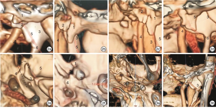
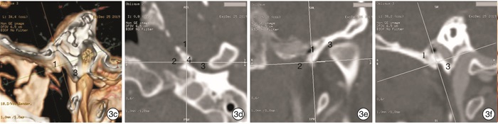
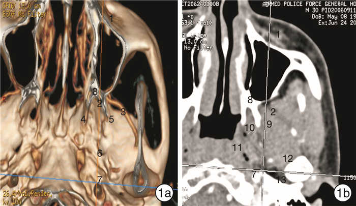
摘要: 目的 探讨咽旁段颈内动脉周边骨性标志的形态及其毗邻关系。方法 随机选取30例(60侧)经64层螺旋CT血管成像(CTA)检查显示颅底结构正常者的影像学资料,由上颌窦入路,观察咽旁段颈内动脉至颈内动脉入口的周边骨性标志,如蝶棘、颞骨鞘突(VPTB)、颞骨鼓嵴(TC)的形态及与咽旁段颈内动脉的毗邻关系。结果 咽旁段颈内动脉,以颈内动脉外口为基准点,分为偏向寰椎方向10侧(16.7%),偏向颞下颌关节方向6侧(10%),靠近正中线44侧(73.3%)。蝶棘的形态53侧(88.3%)成上宽下窄的三角型,6侧(10%)成末端分叉的不规则型,1侧(1.7%)蝶棘缺失。颞骨鞘突形态为扁平的不规则多边形,表面较光滑,60侧(100%)未见缺失。TC是VPTB向蝶棘方向的延续,位于两者之间,其中51侧(85%)存在TC,9侧(15%)TC缺失。三者组成特殊的解剖区域位于咽旁段颈内动脉前外方,可分为反转“J”型43侧(71.7%)、不规则型17侧(28.3%)。结论 蝶棘、TC、VPTB三者构成的特殊解剖区域与由上颌窦入路观察的咽旁段颈内动脉关系密切,是内镜手术寻找咽旁段颈内动脉的解剖依据。CTA影像可提供个体化手术依据。Abstract: Objective To investigate the morphology of bone markers around the parapharyngeal internal carotid artery and its adjacent relationship.Methods The imaging date of 30 cases which had normal structure of the skull by CTA were randomly selected. Through maxillary sinus approach, the morphology of peripheral bony landmarks (sphenoid spine, vaginal process of the tympanic bone, tympanic crest) from the parapharyngeal internal carotid artery to the entrance of the internal carotid artery and the adjacent relationship with the parapharyngeal internal carotid artery were observed for imaging anatomy.Results With the external opening of the internal carotid artery as the reference point, the parapharyngeal internal carotid artery was divided into 10 sides (16.7%) in the direction of atlas and 6 sides(10%) in the direction of temporomandibular joint, 44 sides (73.3%) were close to midline. On 53 sides (88.3%), the morphology of sphenoid spine became triangular shape with upper width and lower width, 6 sides (10%) became irregular type with terminal bifurcation, and 1 side (1.7%) had sphenoid spine missing. The shape of the vaginal process of the tympanic bone (VPTB) was a flat, irregular polygon with a smooth surface and no loss on 60 sides (100%). The tympanic crest (TC) was a continuation of the VPTB to the sphenoid spine. It was located between the sphenoid spine and the VPTB. 51 sides (85%) of the TC and 9 sides (15%) of the TC were missing. The three constituted a special anatomical structure located in front of the internal carotid artery of the parapharyngeal segment, which can be divided into 43 inverted J-types (71.7%) and 17 irregular types (28.3%).Conclusion The special anatomical area composed of sphenoid spine, vaginal process of the tympanic bone, tympanic crest is closely related to the parapharyngeal internal carotid artery observed by maxillary sinus approach, which is the anatomical basis for endoscopic surgery to find the parapharyngeal internal carotid artery. CTA can provide individualized surgical basis.
-

-
[1] 许庚. 经鼻内镜鼻腔鼻窦-颅底肿瘤手术[J]. 中华耳鼻咽喉头颈外科杂志, 2020, 55(1): 4-7. https://www.cnki.com.cn/Article/CJFDTOTAL-SDYU201702002.htm
[2] Falcon RT, Rivera-Serrano CM, Miranda JF, et al. Endoscopic endonasal dissection of the infratemporal fossa: Anatomic relationships and importance of eustachian tube in the endoscopic skull base surgery[J]. Laryngoscope, 2011, 121(1): 31-41. doi: 10.1002/lary.21341
[3] Li W, Chae R, Rubio RR, et al. Characterization of Anatomical Landmarks for Exposing the Internal Carotid Artery in the Infratemporal Fossa Through an Endoscopic Transmasticator Approach: A Morphometric Cadaveric Study[J]. World Neurosurg, 2019, 131: e415-e424. doi: 10.1016/j.wneu.2019.07.185
[4] 王振霖, 张秋航, 刘俊其, 等. 基于解剖平面的内镜经口入路咽旁段颈内动脉定位方法及临床应用[J]. 解剖学报, 2020, 51(5): 677-681. https://www.cnki.com.cn/Article/CJFDTOTAL-JPXB202005010.htm
[5] Kassam AB, Gardner P, Snyderman C, et al. Expanded endonasal approach: fully endoscopic, completely transnasal approach to the middle third of the clivus, petrous bone, middle cranial fossa, and infratemporal fossa[J]. Neurosurg Focus, 2005, 19(1): E6.
[6] Gardner PA, Tormenti MJ, Pant H, et al. Carotid artery injury during endoscopic endonasal skull base surgery: incidence and outcomes[J]. Neurosurgery, 2013, 73(2 Suppl Operative): ons261-269;discussion ons269-270.
[7] Liu J, Pinheiro-Neto CD, Fernandez-Miranda JC, et al. Eustachian tube and internal carotid artery in skull base surgery: an anatomical study[J]. Laryngoscope, 2014, 124(12): 2655-2664. doi: 10.1002/lary.24808
[8] Komune N, Komune S, Matsushima K, et al. Comparison of lateral microsurgical preauricular and anterior endoscopic approaches to the jugular foramen[J]. J Laryngol Otol, 2015, 129 Suppl 2: S12-20.
[9] Rowan NR, Turner MT, Valappil B, et al. Injury of the Carotid Artery during Endoscopic Endonasal Surgery: Surveys of Skull Base Surgeons[J]. J Neurol Surg B Skull Base, 2018, 79(3): 302-308. doi: 10.1055/s-0037-1607314
[10] Kassam AB, Prevedello DM, Carrau RL, et al. Endoscopic endonasal skull base surgery: analysis of complications in the authors' initial 800 patients[J]. J Neurosurg, 2011, 114(6): 1544-1568. doi: 10.3171/2010.10.JNS09406
[11] Labib MA, Prevedello DM, Carrau R, et al. A road map to the internal carotid artery in expanded endoscopic endonasal approaches to the ventral cranial base[J]. Neurosurgery, 2014, 10 Suppl 3: 448-471.
[12] Bouthillier A, van Loveren HR, Keller JT. Segments of the internal carotid artery: a new classification[J]. Neurosurgery, 1996, 38(3): 425-433.
-
 下载:
下载:
图(3)
计量
- 文章访问数: 1451
- PDF下载数: 589
- 施引文献: 0