-
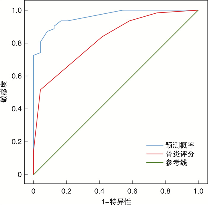
摘要: 目的 探讨单侧上颌窦真菌球(UMFB)的病变特点以提高临床诊断的准确性。方法 回顾性分析2017年1月-2019年6月于山东大学齐鲁医院青岛院区行鼻内镜手术治疗的单侧上颌窦病变且术后经病理证实为UMFB或单侧慢性上颌窦炎(UCMS)患者共86例, 分别从年龄、性别、是否合并糖尿病、病变上颌窦CT特征及上颌窦后外侧壁GOSS骨炎评分进行对比分析, 比较两组病变的差异。CT特征包括: ①上颌窦内见高密度影; ②上颌窦内充满软组织密度影(包括凸入或不凸入中鼻道内软组织密度影); ③上颌窦内未充满软组织密度影(包括表面不平凸起, 表明光滑凸起如类圆形光滑凸起、气泡症)。行卡方、独立样本t检验, Mann-Whitney U检验对差异有统计学意义的指标进行二元Logistic回归分析, 对有意义的数值型变量指标进行受试者工作特征(ROC)曲线分析, 分析分界值与敏感度和特异性的关系, 找到诊断UMFB的最佳截断值。结果 86例单侧上颌窦病变中UMFB 62例(72.1%), UCMS 24例(27.9%)。UMFB好发于中老年患者, 女性多于男性。是否合并糖尿病在两种病变间差异无统计学意义。在鼻窦CT特征上, 经组间比较及二元Logistic回归分析, 上颌窦内高密度影或钙化、上颌窦内未充满软组织密度影表面不平凸起、上颌窦内充满软组织密度影并凸入中鼻道是UMFB的显著预测因素(均P < 0.05)。UMFB骨炎评分明显高于UCMS(P < 0.001), 经ROC曲线分析, 当截断值>3.5时(曲线下面积为0.824), 相应的敏感度和特异性分别为0.516、0.958。结论 通过年龄、性别、CT特征表现以及上颌窦骨炎评分可以将UMFB与单侧上颌窦慢性炎症进行鉴别, 提高临床诊断的准确性。Abstract: Objective The aim of this study is to investigate the pathological characteristics of unilateralmaxillary sinus fungus ball(UMFB) in order to improve the accuracy of clinical diagnosis.Methods A total of 86 patients with unilateral maxillary sinus lesions who underwent nasal endoscopic surgery in Qilu Hospital of Shandong University(Qingdao) from January 2017 to June 2019 were included. Those patients were confirmed UMFB or unilateral chronic maxillary sinusitis(UCMS) by pathology. The characteristics including age, sex, diabetes mellitus or no, CT features of the diseased maxillary sinus and GOSS osteitis score of the posterolateral wall of the maxillary sinus were analyzed, and the differences between the two groups were compared. CT features include: ①intralesional hyperdensity(calcification); ②maxillary sinus full haziness with or without mass effect; ③the irregular lobulated protruding lesion(fuzzy appearance) or smooth. Chi-square, independent sample t test and Mann-Whitney U test were performed. Logistic regression analysis and receiver operating Characteristic(ROC) curve analysis were used to find the best cutoff value for UMFB diagnosis.Results Among the 86 cases of unilateral maxillary sinus lesions, 62 cases were UMFB, which accounted for 72.1%, and 24 cases were UCMS, which accounted for 27.9%. UMFB usually occurs in middle-aged and elderly patients, and there are more females than males. There was no statistical difference between the two groups with or without diabetes. In terms of CT characteristics of paranasal sinuses, intergroup comparison and binary Logistic regression analysis, intralesional hyperdensity, maxillary sinus full haziness with mass effect, the irregular lobulated protruding lesion(fuzzy appearance) were significant predictors of MFB(all P < 0.05). The score of osteitis in UMFB was significantly higher than that in UCMS(P < 0.001). ROC curve analysis showed that when the cutoff value was more than 3.5(the area under the curve was 0.824), the corresponding sensitivity and specificity were 0.516 and 0.958, respectively.Conclusion The age, gender, CT characteristics and maxillary sinus osteitis score can distinguish UMFB from unilateral maxillary sinus chronic inflammation, and improve the accuracy of clinical diagnosis.
-
Key words:
- maxillary sinus /
- sinusitis /
- fungus ball
-

-
表 1 UMFB组与UCMS组上颌窦CT特征比较
指标 UMFB (62例) UCMS (24例) P 高密度影或钙化 44 1 < 0.001 充满上颌窦但未凸入中鼻道 6 6 0.066 充满上颌窦且凸入中鼻道 28 4 0.024 表面不平凸起 22 2 0.012 表面光滑凸起 6 12 < 0.001 GOSS骨炎评分 4[3;4] 2[0.25;3.0] < 0.001 表 2 各项预诊断UMFB指标二元Logistic回归分析结果
预测诊断指标 P OR 95%CI 高密度影或钙化 0.008 71.119 3.038~1665.134 表面不平凸起 0.014 25.357 1.908~337.079 充满上颌窦且凸入中鼻道 0.028 10.154 1.277~80.716 骨炎评分 0.005 7.499 1.824~30.836 -
[1] Liu X, Liu C, Wei H, et al. A retrospective analysis of 1, 717 paranasal sinus fungus ball cases from 2008 to 2017[J]. Laryngoscope, 2020, 130(1): 75-79. doi: 10.1002/lary.27869
[2] 王明婕, 周兵, 李云川, 等. 449例真菌球性鼻窦炎临床特征分析[J]. 临床耳鼻咽喉头颈外科杂志, 2018, 32(3): 220-224. https://www.cnki.com.cn/Article/CJFDTOTAL-LCEH201803017.htm
[3] Costa F, Emanuelli E, Franz L, et al. Fungus ball of the maxillary sinus: Retrospective study of 48 patients and review of the literature[J]. Am J Otolaryngol, 2019, 40(5): 700-704. doi: 10.1016/j.amjoto.2019.06.006
[4] Georgalas C, Videler W, Freling N, et al. Global Osteitis Scoring Scale and chronic rhinosinusitis: a marker of revision surgery[J]. Clin Otolaryngol, 2010, 35(6): 455-461. doi: 10.1111/j.1749-4486.2010.02218.x
[5] Kim JS, So SS, Kwon SH. The increasing incidence of paranasal sinus fungus ball: a retrospective cohort study in two hundred forty-five patients for fifteen years[J]. Clin Otolaryngol, 2017, 42(1): 175-179. doi: 10.1111/coa.12588
[6] Grosjean P, Weber R. Fungus balls of the paranasal sinuses: a review[J]. Eur Arch Otorhinolaryngol, 2007, 264(5): 461-470. doi: 10.1007/s00405-007-0281-5
[7] Lop-Gros J, Gras-Cabrerizo JR, Bothe-González C, et al. Fungus ball of the paranasal sinuses: Analysis of our serie of patients[J]. Acta Otorrinolaringol Esp, 2016, 67(4): 220-225. doi: 10.1016/j.otorri.2015.09.005
[8] 崔昕燕, 汪李琴, 殷敏, 等. 成人单侧鼻-鼻窦病变376例临床病例分析[J]. 临床耳鼻咽喉头颈外科杂志, 2018, 32(6): 439-442, 446. https://www.cnki.com.cn/Article/CJFDTOTAL-LCEH201806010.htm
[9] Yoon YH, Xu J, Park SK, et al. A retrospective analysis of 538 sinonasal fungus ball cases treated at a single tertiary medical center in Korea(1996-2015)[J]. Int Forum Allergy Rhinol, 2017, 7(11): 1070-1075. doi: 10.1002/alr.22007
[10] Seo YJ, Kim J, Kim K, et al. Radiologic characteristics of sinonasal fungus ball: an analysis of 119 cases[J]. Acta Radiol, 2011, 52(7): 790-795. doi: 10.1258/ar.2011.110021
[11] Chen JC, Ho CY. The significance of computed tomographic findings in the diagnosis of fungus ball in the paranasal sinuses[J]. Am J Rhinol Allergy, 2012, 26(2): 117-119. doi: 10.2500/ajra.2012.26.3707
[12] Ho CF, Lee TJ, Wu PW, et al. Diagnosis of a maxillary sinus fungus ball without intralesional hyperdensity on computed tomography[J]. Laryngoscope, 2019, 129(5): 1041-1045. doi: 10.1002/lary.27670
[13] Grosjean P, Weber R. Fungus balls of the paranasal sinuses: a review[J]. Eur Arch Otorhinolaryngol, 2007, 264(5): 461-470. doi: 10.1007/s00405-007-0281-5
[14] Ledderose GJ, Braun T, Betz CS, et al. Functional endoscopic surgery of paranasal fungus ball: clinical outcome, patient benefit and health-related quality of life[J]. Eur Arch Otorhinolaryngol, 2012, 269(10): 2203-2208. doi: 10.1007/s00405-012-1925-7
[15] Kim SC, Ryoo I, Shin JM, et al. MR Findings of Fungus Ball: Significance of High Signal Intensity on T1-Weighted Images[J]. J Korean Med Sci, 2020, 35(3): e22. doi: 10.3346/jkms.2020.35.e22
[16] Zhang J, Li Y, Lu X, et al. Analysis of fungal ball rhinosinusitis by culturing fungal clumps under endoscopic surgery[J]. Int J Clin Exp Med, 2015, 8(4): 5925-5930.
[17] Bosi GR, de Braga GL, de Almeida TS, et al. Fungus ball of the paranasal sinuses: Report of two cases and literature review[J]. Int Arch Otorhinolaryngol, 2012, 16(2): 286-290. doi: 10.7162/S1809-97772012000200020
[18] Dufour X, Kauffmann-Lacroix C, Ferrie JC, et al. Paranasal sinus fungus ball and surgery: a review of 175 cases[J]. Rhinology, 2005, 43(1): 34-39.
[19] Wilson D, Citiulo F, Hube B. Zinc exploitation by pathogenic fungi[J]. PLoS Pathog, 2012, 8(12): e1003034. doi: 10.1371/journal.ppat.1003034
[20] Eide DJ. Homeostatic and adaptive responses to zinc deficiency in Saccharomyces cerevisiae[J]. J Biol Chem, 2009, 284(28): 18565-18569. doi: 10.1074/jbc.R900014200
[21] 邹华, 田鹏. 慢性鼻-鼻窦炎与骨炎[J]. 临床耳鼻咽喉头颈外科杂志, 2015, 29(9): 773-777. https://www.cnki.com.cn/Article/CJFDTOTAL-LCEH201509001.htm
[22] Jun YJ, Shin JM, Lee JY, et al. Bony Changes in a Unilateral Maxillary Sinus Fungal Ball[J]. J Craniofac Surg, 2018, 29(1): e44-e47. doi: 10.1097/SCS.0000000000004010
[23] Tomazic PV, Dostal E, Magyar M, et al. Potential correlations of dentogenic factors to the development of clinically verified fungus balls: A retrospective computed tomography-based analysis[J]. Laryngoscope, 2016, 126(1): 39-43. doi: 10.1002/lary.25416
[24] Mensi M, Salgarello S, Pinsi G, et al. Mycetoma of the maxillary sinus: endodontic and microbiological correlations[J]. Oral Surg Oral Med Oral Pathol Oral Radiol Endod, 2004, 98(1): 119-123. doi: 10.1016/j.tripleo.2003.12.035
[25] 颜旭东, 李娜, 刘培. 真菌球型鼻窦炎合并鼻窦异物1例[J]. 临床耳鼻咽喉头颈外科杂志, 2015, 29(15): 1385-1386. https://www.cnki.com.cn/Article/CJFDTOTAL-LCEH201515020.htm
-

| 引用本文: | 王磊, 袁英, 于学民, 等. 单侧上颌窦真菌球临床特点分析[J]. 临床耳鼻咽喉头颈外科杂志, 2021, 35(4): 328-332. doi: 10.13201/j.issn.2096-7993.2021.04.010 |
| Citation: | WANG Lei, YUAN Ying, YU Xuemin, et al. Analysis of clinical characteristics of fungal ball in unilateral maxillary sinus[J]. J Clin Otorhinolaryngol Head Neck Surg, 2021, 35(4): 328-332. doi: 10.13201/j.issn.2096-7993.2021.04.010 |
- Figure 1.
- Figure 2.
- Figure 3.
- Figure 4.


 下载:
下载:


