
-
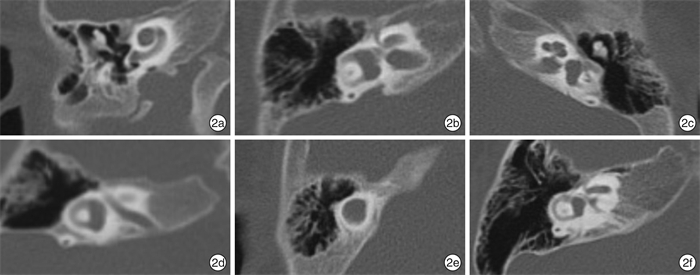
摘要: 目的 分析常见内耳及内听道畸形在单侧聋(SSD)患儿人群中的分布情况,并通过比较SSD患儿聋耳和听力正常耳关键骨性结构参数值,初步研究SSD患儿的影像学病因。方法 收集诊断为SSD且行颞骨高分辨计算机薄层扫描(HRCT)检查的患儿40例,对其进行颞骨HRCT阅片,并测量轴位骨岛面积、前庭宽度、内听道宽度,耳蜗高度及耳蜗底轴宽度,行配对t检验比较SSD患儿聋耳和听力正常耳测量值的差异。结果 SSD聋耳组内耳及内听道畸形率高达62.5%(25/40),最常见的畸形为蜗神经管畸形,其中蜗神经管狭窄20例(50.0%)、蜗神经管闭锁3例(7.5%);其次常见的畸形为内听道畸形,其中内听道狭窄5例(12.5%)、内听道闭锁1例(2.5%);耳蜗畸形有原始听囊畸形1例(2.5%)、不完全分隔(IP)Ⅱ型畸形2例(5.0%)及前庭水管扩大(EVA)畸形1例(2.5%)。听力正常耳组中有1例EVA畸形和1例IP-Ⅱ畸形。聋耳组中内耳各关键结构测量结果相比于听力正常耳组,蜗神经管宽度、内听道宽度及骨岛面积差异有统计学意义(P < 0.05)。除此之外,各项内耳关键结构测量结果差异均无统计学意义。结论 蜗神经管狭窄和内听道狭窄是SSD患儿的主要影像学病因,颞骨HRCT对SSD患儿的影像学病因有较高的诊断价值。Abstract: Objective To investigate the distribution of common inner ear and internal auditory canal malformations in children with single-sided deafness(SSD), and to explore the imaging etiology of SSD by comparing the quantitative parameters of key bone structures between deaf and normal ears in children with congenital SSD. Method Forty children with SSD diagnosed in the Second Hospital of Lanzhou University from September 2016 to March 2019 were collected. All of them underwent HRCT examinations of temporal bone. The area of bone island, the width of vestibular, the width of internal auditory canal, the height of cochlear and the width of cochlear basal axis were measured. Paired t test was used to compare the difference between the hearing abnormality and normal hearing in children with SSD. Result The rate of inner ear deformity was 62.5% in SSD group, the most common deformity was cochlear nerve canal deformity, 20 cases (50.0%) of cochlear canal stenosis and 3 cases (7.5%) of cochlear canal atresia.The second most common deformity was internal auditory canal deformity, including 5 cases (12.5%) of internal auditory canal stenosis and 1 case (2.5%) of internal auditory canal atresia. Other malformations included 1 case(2.5%) of RO, 2 cases (5.0%) of incomplete partition (IP) type II and 1 case (2.5%) of enlargement of vestibular aqueduct (EVA). There are no significant difference in the measured results of the key structures of the inner ear between two groups except the width of cochlear nerve canal, internal auditory canal and the area of bone island. Conclusion The main inner ear deformities in children with SSD are cochlear nerve canal stenosis and inner auditory canal stenosis. HRCT of temporal bone has high diagnostic value for inner ear deformities in children with SSD.
-

-
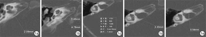
表 1 两位医师测量内耳关键结构的结果一致性比较
x±s 项目 聋耳组 听力正常耳组 医师1 医师2 ICC 医师1 医师2 ICC 蜗神经管宽度/mm 1.37±1.67 1.36±0.72 0.954 2.21±0.34 2.25±0.34 0.881 耳蜗底轴宽度/mm 8.19±1.38 8.22±1.39 0.985 8.42±0.42 8.48±0.43 0.826 耳蜗高度/mm 4.27±0.78 4.13±0.76 0.970 4.40±0.42 4.40±0.47 0.926 骨岛面积/mm2 7.56±2.61 7.05±2.53 0.965 8.09±2.03 7.75±2.07 0.946 前庭宽度/mm 2.88±0.63 3.00±0.66 0.922 3.02±0.43 3.05±0.46 0.808 内听道宽度/mm 3.33±1.05 3.59±1.15 0.988 3.82±0.61 4.12±0.72 0.930  下载: 导出CSV
表 2 聋耳组和听力正常耳组内耳各项测量值比较
x±s 项目 聋耳组 听力正常耳组 t P 蜗神经管宽度/mm 1.35±0.71 2.23±0.32 8.256 0.000 耳蜗底轴宽度/mm 8.20±1.38 8.45±0.39 1.159 0.253 耳蜗高度/mm 4.20±0.71 4.40±0.43 1.920 0.062 骨岛面积/mm2 7.31±2.52 7.92±2.00 2.707 0.010 前庭宽度/mm 2.94±0.63 3.03±0.40 0.992 0.327 内听道宽度/mm 3.46±1.09 3.97±0.64 4.206 0.000
下载: 导出CSV
表 3 40例SSD患儿的内耳及内听道畸形分布情况
畸形 聋耳单侧 双侧 所占比例/% 耳蜗畸形 RO 1 0 2.5 IP-Ⅱ 1 1 5.0 EVA 0 1 2.5 内听道畸形 狭窄 5 0 12.5 闭锁 1 0 2.5 蜗神经管 狭窄 20 0 50.0 闭锁 3 0 7.5
下载: 导出CSV
-
[1] Valente M. Executive Summary: Evidence-Based Best Practice Guideline for Adult Patients with Severe-to-Profound Unilateral Sensorineural Hearing Loss[J]. J Am Acad Audiol, 2015, 26(7): 605-606. doi: 10.3766/jaaa.26.7.2
[2] 石颖, 李永新. 单侧耳聋患者植入相关研究进展[J]. 临床耳鼻咽喉头颈外科杂志, 2017, 31(12): 971-976. https://www.cnki.com.cn/Article/CJFDTOTAL-LCEH201712022.htm
[3] Zeitler DM, Sladen DP, DeJong MD, et al. Cochlear implantation for single-sided deafness in children and adolescents[J]. Int J Pediatr Otorhinolaryngol, 2019, 118: 128-133. doi: 10.1016/j.ijporl.2018.12.037
[4] Usami SI, Kitoh R, Moteki H, et al. Etiology of single-sided deafness and asymmetrical hearing loss[J]. Acta Otolaryngol, 2017, 137(sup565): S2-S7. doi: 10.1080/00016489.2017.1300321
[5] 吴莉, 江杰, 谢晓洁, 等. 先天性感音神经性耳聋HRCT内耳关键性结构定量测量及意义[J]. 中华耳科学杂志, 2018, 16(5): 598-603. doi: 10.3969/j.issn.1672-2922.2018.05.003
[6] Sennaroglu L. Cochlear implantation in inner ear malformations--a review article[J]. Cochlear Implants Int, 2010, 11(1): 4-41. doi: 10.1002/cii.416
[7] Sennaro lu L, Bajin MD. Classification and Current Management of Inner Ear Malformations[J]. Balkan Med J, 2017, 34(5): 397-411. doi: 10.4274/balkanmedj.2017.0367
[8] Song JJ, Choi HG, Oh SH, et al. Unilateral sensorineural hearing loss in children: the importance of temporal bone computed tomography and audiometric follow-up[J]. Otol Neurotol, 2009, 30(5): 604-608. doi: 10.1097/MAO.0b013e3181ab9185
[9] Jackler RK, Luxford WM, House WF. Congenital malformations of the inner ear: a classification based on embryogenesis[J]. Laryngoscope, 1987, 97(3 Pt 2 Suppl 40): 2-14.
[10] Fatterpekar GM, Mukherji SK, Alley J, et al. Hypoplasia of the bony canal for the cochlear nerve in patients with congenital sensorineural hearing loss: initial observations[J]. Radiology, 2000, 215(1): 243-246. doi: 10.1148/radiology.215.1.r00ap36243
[11] Bess FH, Dodd-Murphy J, Parker RA. Children with minimal sensorineural hearing loss: prevalence, educational performance, and functional status[J]. Ear Hear, 1998, 19(5): 339-354. doi: 10.1097/00003446-199810000-00001
[12] Fitzpatrick EM, Al-Essa RS, Whittingham J, et al. Characteristics of children with unilateral hearing loss[J]. Int J Audiol, 2017, 56(11): 819-828. doi: 10.1080/14992027.2017.1337938
[13] Masuda S, Usui S. Comparison of the prevalence and features of inner ear malformations in congenital unilateral and bilateral hearing loss[J]. Int J Pediatr Otorhinolaryngol, 2019, 125: 92-97. doi: 10.1016/j.ijporl.2019.06.028
[14] Shah J, Pham G N, Zhang J, et al. Evaluating diagnostic yield of computed tomography(CT)and magnetic resonance imaging(MRI)in pediatric unilateral sensorineural hearing loss[J]. Int J Pediatr Otorhinolaryngol, 2018, 115: 41-44. doi: 10.1016/j.ijporl.2018.09.003
[15] Chen J X, Kachniarz B, Shin J J. Diagnostic yield of computed tomography scan for pediatric hearing loss: a systematic review[J]. Otolaryngol Head Neck Surg, 2014, 151(5): 718-739. doi: 10.1177/0194599814545727
[16] Ketterer MC, Aschendorff A, Arndt S, et al. The influence of cochlear morphology on the final electrode array position[J]. Eur Arch Otorhinolaryngol, 2018, 275(2): 385-394. doi: 10.1007/s00405-017-4842-y
[17] Miyasaka M, Nosaka S, Morimoto N, et al. CT and MR imaging for pediatric cochlear implantation: emphasis on the relationship between the cochlear nerve canal and the cochlear nerve[J]. Pediatr Radiol, 2010, 40(9): 1509-1516. doi: 10.1007/s00247-010-1609-7
[18] 燕飞, 李建红, 李静, 等. 儿童蜗神经发育不良的影像学表现[J]. 放射学实践, 2011, 26(3): 260-263. doi: 10.3969/j.issn.1000-0313.2011.03.009
[19] Friedman AB, Guillory R, Ramakrishnaiah R H, et al. Risk analysis of unilateral severe-to-profound sensorineural hearing loss in children[J]. Int J Pediatr Otorhinolaryngol, 2013, 77(7): 1128-1131. doi: 10.1016/j.ijporl.2013.04.016
[20] Uwiera TC, DeAlarcon A, Meinzen-Derr J, et al. Hearing loss progression and contralateral involvement in children with unilateral sensorineural hearing loss[J]. Ann Otol Rhinol Laryngol, 2009, 118(11): 781-785. doi: 10.1177/000348940911801106
[21] Marcus S, Whitlow CT, Koonce J, et al. Computed tomography demonstrates abnormalities of contralateral ear in subjects with unilateral sensorineural hearing loss[J]. Int J Pediatr Otorhinolaryngol, 2014, 78(2): 268-271. doi: 10.1016/j.ijporl.2013.11.020
-
图(2)
表(3)
计量
- 文章访问数: 1280
- PDF下载数: 258
- 施引文献: 0