Application of 3D U-net in automatic segmentation of middle ear surgery structures in temporal bone CT
-
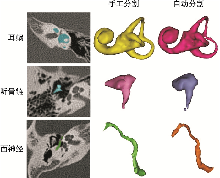
摘要: 目的 研究基于三维卷积神经网络3D U-net神经网络的深度学习方法, 在临床常规颞骨CT影像中, 对中耳手术相关的迷路、面神经和听骨进行全自动分割的可行性。方法 随机调取门诊常规行颞骨CT检查患者的薄层CT扫描数据30例为正常结构组, 既往耳蜗、面神经和听骨形态或走行变异者各1例为异常结构组。所有数据由两位临床医生在Mimics 20.0软件中, 对面神经、迷路和听骨3个结构进行手工初分割和精细分割, 同时利用3D U-Net对上述数据进行深度学习。分别对正常结构组中测试集5例和异常结构组中的迷路、听骨和面神经进行手工分割与自动分割的Dice相似指数(DSC)比较。结果 利用3D U-net网络结构对常规颞骨CT中迷路、听骨和面神经进行自动分割, 其DSC分别为0.79±0.03、0.64±0.05和0.49±0.09;对异常的迷路、听骨和面神经的识别, 其DSC也可达到0.71、0.54和0.40。结论 根据颞骨解剖特点, 采用3D U-net神经网络结构, 可以实现对迷路、听骨和面神经的全自动化分割, 并获得接近手工分割的精度, 该方法可行、快捷、准确度高。Abstract: Objective To study the feasibility of fully automatic segmentation of labyrinth, facial nerve and ossicles in clinical routine temporal bone CT images based on 3D U-net neural network. Method Clinical data were divided into two groups: ①Normal group: data were randomly assigned from 30 patients for routine temporal bone CT examination; ②Abnormal group: cochlear, ossicles and facial nerve morphology variation of 1 case each. The structures of facial nerve, labyrinth and ossicles were manually initial segmented and fine segmented by 2 clinicians with Mimics 20.0. Three-dimensional convolutional neural network(3D U-Net) was selected to conduct deep learning on the same data. The dice similarity coefficient(DSC) was used as the evaluation index. Result The 3D U-net neural network was used to automatically segment the labyrinth, ossicles and facial nerve in the routine temporal bone CT. In the normal group, the DSC of labyrinth, ossicles and facial nerve were 0.79±0.03, 0.64±0.05 and 0.49±0.09, respectively. In the abnormal group, the DSC of these structures were 0.71, 0.54 and 0.40.Conclusion According to the anatomical characteristics of the temporal bone, the labyrinth, ossicles and the facial nerve can be totally automatic segmented by 3D U-net neural network, and the accuracy was closed to that of manual segmentation. This method is feasible, fast and accurate.
-

-
表 1 自动分割与手工分割的DSC比较
例数 迷路 听骨 面神经 正常结构组 训练集 1 250 0.81±0.02 0.63±0.05 0.54±0.06 测试集 5 0.79±0.03 0.64±0.05 0.49±0.09 异常结构组 1 0.71 0.54 0.40 -
[1] 潘亚玲, 王晗琦, 陆勇. 人工智能在医学影像CAD中的应用[J]. 国际医学放射学杂志, 2019, 42(1): 3-7. https://www.cnki.com.cn/Article/CJFDTOTAL-GWLC201901002.htm
[2] 夏黎明, 沈坚, 张荣国, 等. 深度学习技术在医学影像领域的应用[J]. 协和医学杂志, 2018, 9(1): 10-14. doi: 10.3969/j.issn.1674-9081.2018.01.003
[3] 宫进昌, 赵尚义, 王远军. 基于深度学习的医学图像分割研究进展[J]. 中国医学物理学杂志, 2019, 36(4): 420-424. doi: 10.3969/j.issn.1005-202X.2019.04.010
[4] Wullstein H. Theory and practice of tympanoplasty[J]. Laryngoscope, 1956, 66(8): 1076-1093.
[5] Fauser J, Stenin I, Bauer M, et al. Toward an automatic preoperative pipeline for image-guided temporal bone surgery[J]. Int J Comput Assist Radiol Surg, 2019, 14(6): 967-976. doi: 10.1007/s11548-019-01937-x
[6] Elfarnawany M, Rohani SA, Ghomashchi S, et al. Improved middle-ear soft-tissue visualization using synchrotron radiation phase-contrast imaging[J]. Hear Res, 2017, 354: 1-8. doi: 10.1016/j.heares.2017.08.001
[7] Powell KA, Liang T, Hittle B, et al. Atlas-based segmentation of temporal bone anatomy[J]. Int J Comput Assist Radiol Surg, 2017, 12(11): 1937-1944. doi: 10.1007/s11548-017-1658-6
[8] 马晓波, 赵守琴, 李洁, 等. 正常耳颞骨内面神经形态分析[J]. 中国耳鼻咽喉头颈外科, 2015, 22(6): 287-289. https://www.cnki.com.cn/Article/CJFDTOTAL-EBYT201506010.htm
[9] Noble JH, Labadie RF, Majdani O, et al. Automatic segmentation of intracochlear anatomy in conventional CT[J]. IEEE Trans Biomed Eng, 2011, 58(9): 2625-2632. doi: 10.1109/TBME.2011.2160262
[10] Reda FA, Noble JH, Rivas A, et al. Automatic segmentation of the facial nerve and chorda tympani in pediatric CT scans[J]. Med Phys, 2011, 38(10): 5590-5600. doi: 10.1118/1.3634048
-


 下载:
下载:
