
Characteristics of resting state functional magnetic resonance imaging in tinnitus patients with different degrees of affective disorder
-
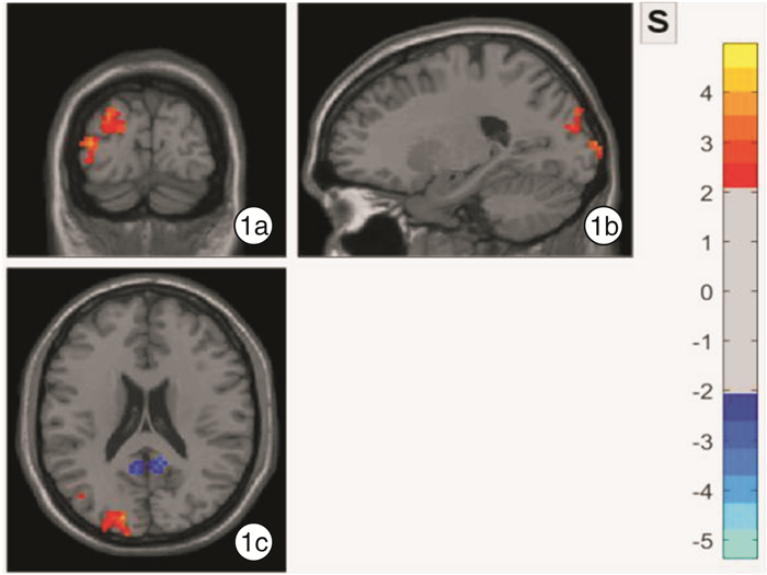
摘要: 目的 观察不同程度情感障碍的耳鸣患者与无耳鸣健康志愿者的静息态功能磁共振成像(rs-fMRI)的差异,分析因耳鸣导致的不同程度情感障碍与脑功能变化的相关性。方法 选取曾参与前期研究的耳鸣患者19例,同时选取同期无耳鸣的健康志愿者15例作为对照。根据耳鸣致残量表得分将患者分为轻度组和重度组。耳鸣患者与健康志愿者均采集rs-fMRI,利用DPABI软件对结果分别进行局部一致性(ReHo)分析、低频波动振幅(ALFF)分析和低频波动分数振幅(fALFF)分析获得ReHo值、ALFF值和fALFF值。将轻度组与对照组、重度组与对照组的ReHo值、ALFF值和fALFF值分别行双样本t检验。结果 与对照组比较,轻度组左枕中回的fALFF值高于对照组,差异有统计学意义(P < 0.05),但ALFF值和ReHo值的差异无统计学意义;重度组右额上回、左颞中回、左额下回三角部和左尾状核的ALFF值明显高于对照组,差异有统计学意义(P < 0.05),但2组fALFF值和ReHo值差异无统计学意义。结论 耳鸣导致的情感障碍程度不同时,脑功能异常区域不同。轻度情感障碍时可通过fALFF分析检出神经活动增高的脑区为左枕中回。重度情感障碍可通过ALFF分析检出神经活动增高的脑区为左颞中回、右额上回、左额下回三角部、左尾状核。
-
关键词:
- 耳鸣 /
- 情感障碍 /
- 静息态功能磁共振成像
Abstract: Objective To analyze the correlation of the degree of affective disorder and brain function changes by comparing the differences of resting-state functional Magnetic Resonance Imaging(rs-fMRI) between healthy volunteers without tinnitus and patients with tinnitus.Method A analysis of 19 patients with tinnitus and 15 healthy volunteers without tinnitus. The patients were divided into mild group and severe group according to tinnitus handicap inventory(THI). Rs-fMRI was collected and the regional homogeneity(ReHo) analysis, amplitude of low-frequency fluctuation(ALFF) analysis, and fractional amplitude of low frequency fluctuation(fALFF) analysis of rs-fMRI were performed by DPABI software. Two-sample t-test of the ReHo value, ALFF value and fALFF value between the mild group and the control group, the severe group and the control group, were performed respectively.Result The fALFF value of the left occipital gyrus in the mild group was higher than that in the control group, the difference was statistically significant(P < 0.05), but there is no statistically significant difference of ALFF value and ReHo value between two groups. The ALFF value of the middle temporal gyrus(left), superior frontal gyrus(right), inferior frontal gyrus pars triangularis(left) and caudate nucleus(left) in the severe group were higher than those of the control group. But there was no significant difference in the fALFF value and the ReHo value.Conclusion Different severity of affective disorder in patients with tinnitus have different areas of brain function abnormalities. Mild group was detected by fALFF analysis and the active brain area was the left middle occipital region. Severe group was detected by ALFF analysis. The active brain regions were left middle temporal gyrus, right superior frontal gyrus, left inferior frontal gyrus pars triangularis, and left caudate nucleus. -

-
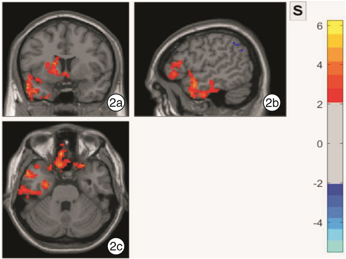
表 1 重度组与对照组比较ALFF值显著增高的脑区
脑区 t值 各维度MNI峰值/mm 簇大小(体素) X Y Z 颞中回(左) 4.8467 -51 6 -24 536 额上回(右) 6.2413 12 60 0 887 额下回三角部(左) 4.2976 -39 42 0 223 尾状核(左) 4.3337 -15 9 12 265  下载: 导出CSV
-
[1] 齐思涵, 秦兆冰, 陈秀兰, 等. 影响慢性主观性耳鸣严重程度的相关因素分析[J]. 听力学及言语疾病杂志, 2014, 22(4): 367-370. doi: 10.3969/j.issn.1006-7299.2014.04.008
[2] 徐建乐, 郑芸, 孟照莉, 等. 耳鸣严重程度与焦虑的相关性研究[J]. 临床耳鼻咽喉头颈外科杂志, 2012, 26(16): 729-732. https://www.cnki.com.cn/Article/CJFDTOTAL-LCEH201216006.htm
[3] 蔡伟伟, 区洁楹, 梁健刚, 等. 耳鸣静息态磁共振局部一致性研究[J]. 实用医学杂志, 2017, 33(14): 2329-2332. doi: 10.3969/j.issn.1006-5725.2017.14.020
[4] 韩祺, 刘代洪, 王尧, 等. 主观耳鸣患者大脑局部神经功能改变的静息态功能磁共振研究[J], 磁共振成像, 2018, 9(7): 481-486. https://www.cnki.com.cn/Article/CJFDTOTAL-CGZC201807003.htm
[5] 石秋兰, 卜行宽, 王俊国, 等. 耳鸣致残量表中文版的研译与临床应用[J]. 南京医科大学学报(自然科学版), 2007, 27(5): 476-479. https://www.cnki.com.cn/Article/CJFDTOTAL-NJYK200705017.htm
[6] Teng C, Zhou J, Ma H, et al. Abnormal resting state activity of left middle occipital gyrus and its functional connectivity in female patients with major depressive disorder[J]. BMC Psychiatry, 2018, 18(1): 370. doi: 10.1186/s12888-018-1955-9
[7] Assaf M, Hyatt CJ, Wong CG, et al. Mentalizing and motivation neural function during social interactions in autism spectrum disorders[J]. Neuroimage Clin, 2013, 3: 321-331. doi: 10.1016/j.nicl.2013.09.005
[8] Cheng C, Dong D, Jiang Y, et al. State-Related Alterations of Spontaneous Neural Activity in Current and Remitted Depression Revealed by Resting-State fMRI[J]. Front Psychol, 2019, 10: 245. doi: 10.3389/fpsyg.2019.00245
[9] Fan J, Zhong M, Gan J, et al. Spontaneous neural activity in the right superior temporal gyrus and left middle temporal gyrus is associated with insight level in obsessive-compulsive disorder[J]. J Affect Disord, 2017, 207: 203-211. doi: 10.1016/j.jad.2016.08.027
[10] Liu CH, Tang LR, Gao Y, et al. Resting-state mapping of neural signatures of vulnerability to depression relapse[J]. J Affect Disord, 2019, 250: 371-379. doi: 10.1016/j.jad.2019.03.022
[11] Ogawa R, Kagitani-Shimono K, Matsuzaki J, et al. Abnormal cortical activation during silent reading in adolescents with autism spectrum disorder[J]. Brain Dev, 2019, 41(3): 234-244. doi: 10.1016/j.braindev.2018.10.013
[12] Wang S, Zhao Y, Zhang L, et al. Stress and the brain: Perceived stress mediates the impact of the superior frontal gyrus spontaneous activity on depressive symptoms in late adolescence[J]. Hum Brain Mapp, 2019, 40(17): 4982-4993. doi: 10.1002/hbm.24752
[13] Roberts G, Lord A, Frankland A, et al. Functional Dysconnection of the Inferior Frontal Gyrus in Young People With Bipolar Disorder or at Genetic High Risk[J]. Biol Psychiatry, 2017, 81(8): 718-727. doi: 10.1016/j.biopsych.2016.08.018
[14] Cha J, DeDora D, Nedic S, et al. Clinically Anxious Individuals Show Disrupted Feedback between Inferior Frontal Gyrus and Prefrontal-Limbic Control Circuit[J]. J Neurosci, 2016, 36(17): 4708-4718. doi: 10.1523/JNEUROSCI.1092-15.2016
[15] Zhang L, Li B, Wang H, et al. Decreased middle temporal gyrus connectivity in the language network in schizophrenia patients with auditory verbal hallucinations[J]. Neurosci Lett, 2017, 653: 177-182. doi: 10.1016/j.neulet.2017.05.042
[16] Perez PL, Wang SS, Heath S, et al. Human caudate nucleus subdivisions in tinnitus modulation[J]. J Neurosurg, 2019: 1-7.
-
图(2)
表(1)
计量
- 文章访问数: 817
- PDF下载数: 640
- 施引文献: 0