
Analysis of the factors for the lymph node metastasis in rⅥb region of thyroid micropapillary carcinoma and the application value of intraoperative nano-carbon
-
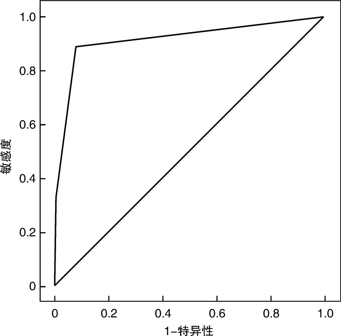
摘要: 目的 探讨rⅥb区淋巴结转移与甲状腺微小乳头状癌病理特征的关系及术中使用纳米碳对手术及rⅥb区淋巴结清扫的应用价值。方法 根据175例甲状腺微小乳头状癌术中是否使用纳米碳,分析纳米碳使用对手术时间、中央区、rⅥb区淋巴结清扫出的数量、术后低钙症状和声嘶情况发生的影响; 将rⅥb区淋巴结转移与否与甲状腺微小乳头状癌病理特征进行统计分析; ROC曲线分析彩超学特征在rⅥb区淋巴结阳性率评估中的价值。结果 甲状腺微小乳头状癌术中使用纳米碳,可提高中央区、rⅥb区淋巴结清扫出的数量,降低甲状旁腺损伤的概率,差异有统计学意义; rⅥb区淋巴结阳性率与年龄、性别、结节纵横比及微小钙化无关,与肿瘤大小、多灶性及是否侵犯被膜有关; 以上述3种彩超相关特征建立rⅥb区淋巴结转移评估模型,ROC曲线分析表明其具有较高的应用价值。结论 肿瘤大小、多灶性及是否侵犯被膜是甲状腺微小乳头状癌rⅥb区淋巴结转移的危险因素,术中使用纳米碳可提高颈部淋巴结清扫的彻底性并增加手术安全性。Abstract: Objective To investigate the relationship between lymph node metastasis in rⅥb region and pathological features of papillary thyroid microcarcinoma, and the application value of carbon nanoparticles in the operation for lymph node dissection in rⅥb region.Method One hundred and seventy-five patients were divided into carbon nanoparticles group and the control group depending on whether carbon nanoparticles were used in the operation. The operation time, the number of central lymph node, the number of rⅥb region lymph node, hypocalcaemia symptom complex and hoarseness after the operaion were compared between the two groups. The lymph node metastasis in the rⅥb region and the pathological features of papillary thyroid microcarcinoma were statistically analyzed.Result The amount of lymph nodes in Central District and rⅥb region was increased and the probability of parathyroid gland injury was decreased by using nano-carbon. The positive rate of lymph nodes in rⅥb region was not related to age, sex, aspect ratio of nodules and microcalcification, but was related to the tumor size, multifoci and the invasion of the capsule. The evaluation model of lymph node metastasis in rⅥb region was established based on the above three correlative features of color doppler ultrasound. The ROC curve analysis showed that the model had high application value.Conclusion The tumor size, multiple foci and capsule invasion are risk factors for lymph node metastasis in rⅥb region of thyroid micropapillary carcinoma.
-

-
表 1 2组手术时间、淋巴结检出数目、术后手足麻木、声嘶情况
组别 例数 手术时间/min 中央区淋巴结个数
(枚/例)rⅥb区淋巴结个数
(枚/例)术后手足麻木
/例术后声嘶
/例纳米碳组 102 131.17±9.28 8.27±1.59 3.28±0.49 6 5 对照组 73 129.25±9.13 5.43±1.42 1.18±0.32 12 11 T(χ2) 0.72 12.24 6.45 5.14 5.29 P 0.57 < 0.01 0.01 0.02 0.02  下载: 导出CSV
表 2 rⅥb区淋巴结阳性率与甲状腺微小乳头状癌病理特征的关系
病理学特征 例数 rⅥb淋巴结 χ2 P (+) (-) 性别 男 37 13 24 0.00 0.97 女 138 48 90 年龄/岁 < 45 89 34 55 0.89 0.35 ≥45 86 27 59 结节大小/mm ≥5 109 49 60 12.98 0.00 < 5 66 12 54 多灶性 单灶 102 15 87 43.73 0.00 多灶 73 46 27 结节纵横比 < 1 51 16 35 0.39 0.54 ≥1 124 45 79 微小钙化 有 127 41 86 1.35 0.25 无 48 20 28 病灶血供丰富 是 34 12 22 0.00 0.95 否 141 49 92 是否侵犯被膜 是 22 15 7 12.31 0.00 否 153 46 107 是否合并桥本 是 32 10 22 0.22 0.64 否 143 51 92 病灶位置 上1/2 105 31 74 3.29 0.07 下1/2 70 30 40
下载: 导出CSV
-
[1] Suzuki S, Bogdanova TI, Saenko VA, et al. Histopathological analysis of papillary thyroid carcinoma detected during ultrasound screening examinations in Fukushima[J]. Cancer Sci, 2019, 110(2): 817-827. doi: 10.1111/cas.13912
[2] Gur EO, Karaisli S, Haciyanli S, et al. Multifocality related factors in papillary thyroid carcinoma[J]. Asian J Surg, 2019, 42(1): 297-302. doi: 10.1016/j.asjsur.2018.05.004
[3] Higashino M, Ayani Y, Terada T, et al. Clinical features of poorly differentiated thyroid papillary carcinoma[J]. Auris Nasus Larynx, 2019, 46(3): 437-442. doi: 10.1016/j.anl.2018.10.001
[4] Lee YM, Park JH, Cho JW, et al. The definition of lymph node micrometastases in pathologic N1a papillary thyroid carcinoma should be revised[J]. Surgery, 2019, 165(3): 652-656. doi: 10.1016/j.surg.2018.09.015
[5] Rosario PW, Mourão G, Calsolari MR. Risk of recurrence in patients with papillary thyroid carcinoma and minimal extrathyroidal extension not treated with radioiodine[J]. J Endocrinol Invest, 2019, 42(6): 687-692. doi: 10.1007/s40618-018-0969-y
[6] Vuong HG, Long NP, Anh NH, et al. Papillary thyroid carcinoma with tall cell features is as aggressive as tall cell variant: a meta-analysis[J]. Endocr Connect, 2018, 7(12): R286-R293. doi: 10.1530/EC-18-0333
[7] Gorostis S, Raguin T, Schneegans O, et al. Incidental thyroid papillary microcarcinoma: survival and follow-up[J]. Laryngoscope, 2019, 129(7): 1722-1726. doi: 10.1002/lary.27664
[8] Zheng KS, Zeng Y, Chen C, et al. Risk factors of cervical lymph node metastasis in papillary thyroid microcarcinoma: an analysis based on data from the surveillance, epidemiology and end results database[J]. Zhongguo Yi Xue Ke Xue Yuan Xue Bao, 2018, 40(6): 736-743.
[9] Henke LE, Pfeifer JD, Baranski TJ, et al. Long-term outcomes of follicular variant vs classic papillary thyroid carcinoma[J]. Endocr Connect, 2018, 7(12): 1226-1235. doi: 10.1530/EC-18-0264
[10] Wang B, Su AP, Xing TF, et al. The function of carbon nanoparticles to improve lymph node dissection and identification of parathyroid glands during thyroid reoperation for carcinoma[J]. Medicine(Baltimore), 2018, 97(32): e11778.
[11] 付浩, 张朝林, 唐振宁, 等. 纳米碳在甲状腺乳头状癌Ⅵ区淋巴结清扫术中应用的研究[J]. 临床耳鼻咽喉头颈外科杂志, 2017, 31(14): 1089-1092. https://www.cnki.com.cn/Article/CJFDTOTAL-LCEH201714009.htm
[12] Yan B, Hou Y, Chen D, et al. Risk factors for contralateral central lymph node metastasis in unilateral cN0 papillary thyroid carcinoma: A meta-analysis[J]. Int J Surg, 2018, 59(1): 90-98.
[13] Gong Y, Yang J, Yan S, et al. Pattern of and clinicopathologic risk factors for lateral lymph node metastases in papillary thyroid carcinoma patients with lateral cervical lymphadenopathy[J]. Medicine(Baltimore), 2018, 97(36): e12263.
[14] Cho JG, Byeon HK, Oh KH, et al. Clinicopathological significance of cancer-associated fibroblasts in papillary thyroid carcinoma: a predictive marker of cervical lymph node metastasis[J]. Eur Arch Otorhinolaryngol, 2018, 275(9): 2355-2361. doi: 10.1007/s00405-018-5061-x
[15] Liu L, Oh C, Heo JH, et al. Clinical significance of extrathyroidal extension according to primary tumor size in papillary thyroid carcinoma[J]. Eur J Surg Oncol, 2018, 44(11): 1754-1759. doi: 10.1016/j.ejso.2018.05.009
[16] Zhang T, Qu Y, He L, et al. Risk factors and preoperative evaluation of lymph nodes posterior to right recurrent laryngeal nerve metastasis in thyroid papillary carcinoma[J]. Zhonghua Yi Xue Za Zhi, 2018, 98(22): 1775-1779.
[17] Yagmur Y, Akbulut S, Sakarya H, et al. Assessment of the relationship between clinical and histopathological features in cases of thyroidectomy[J]. Ann Ital Chir, 2018, 89(2): 199-205.
[18] Yoo RE, Kim JH, Jang EH, et al. Prediction of nondiagnostic results in fine-needle aspiration of thyroid nodules: utility of on-site gross visual assessment of specimens for liquid-based cytology[J]. Endocr Pract, 2018, 24(10): 867-874. doi: 10.4158/EP-2018-0183
[19] Park HK, Kim DW, Ha TK, et al. Utility of routine ultrasonography follow-up after total thyroidectomy in patients with papillary thyroid carcinoma: a single-center study[J]. BMC Med Imaging, 2018, 18(1): 12-12. doi: 10.1186/s12880-018-0253-9
[20] 庞玉娟, 陈晓红, 张景义, 等. 原发灶不明的颈部淋巴结甲状腺乳头状转移癌的临床治疗[J]. 临床耳鼻咽喉头颈外科杂志, 2017, 31(13): 1013-1016. https://www.cnki.com.cn/Article/CJFDTOTAL-LCEH201713011.htm
[21] 乔雷, 董朝, 张楠, 等. 甲状腺乳头状癌淋巴结跳跃转移规律分析[J]. 临床耳鼻咽喉头颈外科杂志, 2018, 32(7): 522-526. https://www.cnki.com.cn/Article/CJFDTOTAL-LCEH201807012.htm
[22] Maksimovic S, Jakovljevic B, Gojkovic Z. Lymph node metastases papillary thyroid carcinoma and their importance in recurrence of disease[J]. Med Arch, 2018, 72(2): 108-111. doi: 10.5455/medarh.2018.72.108-111
[23] Tang T, Li J, Zheng L, et al. Risk factors of central lymph node metastasis in papillary thyroid carcinoma: A retrospective cohort study[J]. Int J Surg, 2018, 54(Pt A): 129-132.
[24] Wang XQ, Wei W, Wei X, et al. Study on the relationship between ultrasonographic features of papillary thyroid carcinoma and central cervical lymph node metastasis[J]. Zhonghua Zhong Liu Za Zhi, 2018, 40(3): 196-200.
[25] Murakami Y, Shimura T, Okada R, et al. Pancreatic metastasis of papillary thyroid carcinoma preoperatively diagnosed by endoscopic ultrasound-guided fine-needle aspiration biopsy: a case report with review of literatures[J]. Clin J Gastroenterol, 2018, 11(6): 521-529. doi: 10.1007/s12328-018-0875-z
[26] Yan S, Zhao W, Wang B, et al. Preoperative injection of carbon nanoparticles is beneficial to the patients with thyroid papillary carcinoma: From a prospective study of 102 cases[J]. Medicine(Baltimore), 2018, 97(27): e11364.
-
图(1)
表(2)
计量
- 文章访问数: 1794
- PDF下载数: 465
- 施引文献: 0