-
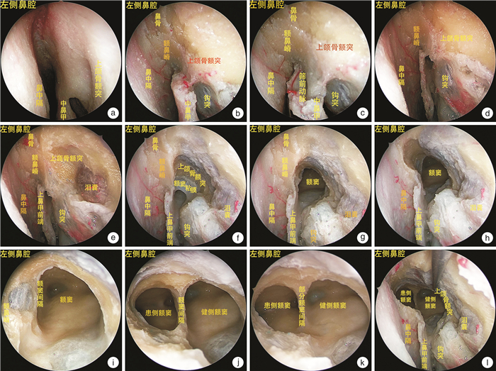
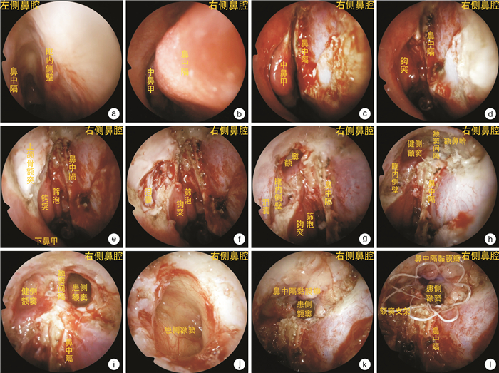
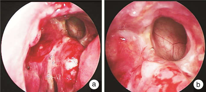
摘要: 目的 患侧额窦的引流通道解剖变异或瘢痕闭锁,经患侧额窦引流通道开放额窦,可能会导致手术失败。本研究拟探索一种借健侧额窦和额隐窝为通路,磨除额窦底壁及额窦间隔,连通双侧额窦的改良DrafⅢ手术术式,完成患侧额窦的引流。方法 通过对2个头颅骨性标本和2个新鲜冷冻标本的解剖研究,探索手术相关标志及手术路径。回顾性分析4例采用此术式进行治疗的患者。记录患者的相关临床资料,探讨该术式的技术细节和优缺点。结果 通过2例头颅解剖研究,确认手术路径,借健侧额窦和额隐窝为通路,磨除双侧额窦底壁和额窦间隔,将双侧额窦在鼻中隔上方扩大成为一个大的共同腔,完成患侧额窦在健侧鼻腔的中线引流。4例患者因单侧额窦乳头状瘤行DrafⅡb手术,术后发生额窦闭锁、额窦炎,遂采用DrafⅢ借道引流术进行治疗。术后患者头痛症状消失,内镜下检查额窦引流口宽敞、黏膜愈合良好、引流通畅,无其他术后并发症。结论 单侧额窦入路DrafⅢ借道引流术能充分引流患侧额窦。该术式创伤小,成功率高,有临床应用价值,适合单侧DrafⅡb手术失败的患者。Abstract: Objective Anatomical variation or scar atresia of the drainage channel of the frontal sinus on the affected side, and opening the frontal sinus through the drainage channel of the frontal sinus on the affected side may lead to surgical failure. The purpose of this study is to explore a modified Draf Ⅲ operation to complete the drainage of the affected frontal sinus by removing the floor wall and septum of the frontal sinus and connecting the bilateral frontal sinus through the healthy side of the frontal sinus.Methods Through the anatomical study of 2 skull bone specimens and 2 fresh frozen specimens, the surgical landmark and surgical approach were explored. Four patients with frontal sinus atresia and frontal sinusitis after DrafⅡb surgery in Eye & ENT Hospital of Fudan University were retrospectively analyzed. Descriptive method was used to analyze the data.Results The bottom wall of bilateral frontal sinus was removed, and the bilateral frontal sinus was enlarged above the nasal septum to form a large common cavity. The uncinate process and ethmoid bubble were retained, and the midline drainage of the affected frontal sinus in the healthy side of the nasal cavity was completed. From August 2022 to April 2023, 4 patients with frontal sinus atresia and frontal sinusitis after DrafⅡb surgery for unilateral frontal sinus papilloma in Eye & ENT Hospital of Fudan University were treated with surgery. The headache symptoms disappeared after surgery, and the drainage of frontal sinus was spacious, the mucosa healed well and the drainage was unobstructed under endoscopy. There were no other postoperative complications.Conclusion DrafⅢ approach to unilateral frontal sinus for contralateral drainage can drain the affected frontal sinus adequately. The essence of this operation is to drain the bilateral frontal sinus in the unilateral nasal cavity, and this operation has short path, less trauma, and a broader prospect, which is suitable for promotion.
-
Key words:
- DrafⅢ /
- maxillary frontal process /
- fronto-nasal ridge /
- unilateral frontal sinus
-

-
表 1 4例内镜单侧额窦入路DrafⅢ借道引流术患者一般资料
例序 性别 年龄/岁 临床诊断 手术方案 术后4周鼻内镜情况 1 女 17 额窦炎(左) 内镜经单(右)侧额窦入路DrafⅢ借道引流术、中鼻甲黏膜瓣修复术 4例患者均额窦口宽敞,额窦引流通畅、未见瘢痕及滤泡形成、中鼻甲黏膜瓣成活良好 2 男 43 额窦炎(左) 内镜经单(右)侧额窦入路DrafⅢ借道引流术、中鼻甲黏膜瓣修复术 3 男 49 额窦炎(右) 内镜经单(左)侧额窦入路DrafⅢ借道引流术、中鼻甲黏膜瓣修复术 4 男 59 额窦炎(右) 内镜经单(左)侧额窦入路DrafⅢ借道引流术、中鼻甲黏膜瓣修复术 -
[1] Draf W. Endonasal microendoscopic frontal sinus surgery: The fulda concept[J]. Oper Tech Otolaryngol Head Neck Surg, 1991, 2(4): 234-240. doi: 10.1016/S1043-1810(10)80087-9
[2] 余洪猛, 郑春泉, 王德辉, 等. DrafⅠ~Ⅲ型鼻内额窦引流术[J]. 临床耳鼻咽喉头颈外科杂志, 2007, 7(21): 664-667. https://www.cnki.com.cn/Article/CJFDTOTAL-LCEH200714020.htm
[3] Zhao KQ, Pu SL, Yu HM. Endoscopic Septoplasty with Limited Two-line Resection: Minimally Invasive Surgery for Septal Deviation[J]. J Vis Exp, 2018, 136: 57678.
[4] 马有祥, 田昊. 中线入路额窦Draf Ⅲ型手术[J]. 中华耳鼻咽喉头颈外科杂志, 2022, 57(8): 910-914. doi: 10.3760/cma.j.cn115330-20220107-00012
[5] Chin D, Snidvongs K, Kalish L, et al. The outsidein approach to the modified endoscopic Lothrop procedure[J]. Laryngoscope, 2012, 122(8): 1661-1669. doi: 10.1002/lary.23319
[6] Nishiike S, Yoda S, Shikina T, et al. Endoscopic Transseptal Approach to Frontal Sinus Disease[J]. Indian J Otolaryngol Head Neck Surg, 2015, 67(3): 287-291. doi: 10.1007/s12070-015-0879-7
[7] Gross CW, Schlosser RJ. The modified Lothrop procedure: lessons learned[J]. Laryngoscope, 2001, 111(7): 1302-1305. doi: 10.1097/00005537-200107000-00030
[8] Wormald PJ. Salvage frontal sinus surgery: The endoscopic modified Lothrop procedure[J]. Laryngoscope, 2003, 113(2): 276-283. doi: 10.1097/00005537-200302000-00015
[9] Kuhn FA. Chronic frontal sinusitis: the endoscopic frontal recess approach[J]. Otolaryngol Head Neck Surg, 1996, 7: 222-229.
[10] Wormald PJ, Hoseman W, Callejas C, et al. The international frontal sinus anatomy classsification(IFCA)and classsification of the extent of endoscopic frontal sinus surgery(FESS)[J]. Int Forum Allergy Rhinol, 2016, 6(7): 677-696. doi: 10.1002/alr.21738
[11] Özer M, Kutlay AM, Durmaz MO, et al. Extended endonasal endoscopic approach for anterior midline skull base lesions[J]. Clin Neurol Neurosurg, 2020, 196: 106024. doi: 10.1016/j.clineuro.2020.106024
[12] Gotlib T, Held-Ziółkowska M, Niemczyk K. Extended draf Ⅱb procedures in the treatment of frontal sinus pathology[J]. Clin Exp Otorhinolaryngol, 2015, 8(1): 34-38. doi: 10.3342/ceo.2015.8.1.34
[13] Lai WS, Yang PL, Lee CH, et al. The association of frontal recess anatomy and mucosal disease on the presence of chronic frontal sinusitis: a computed tomographic analysis[J]. Rhinology, 2014, 52(3): 208-214. doi: 10.4193/Rhino13.110
[14] Gross CW, Schlosser RJ. The modified Lothrop procedure: lessons learned[J]. Laryngoscope, 2001, 111(7): 1302-1305. doi: 10.1097/00005537-200107000-00030
[15] Lee JT, Kennedy DW, Palmer JN, et al. The incidence of concurrent osteitis in patients with chronic rhinosinusitis: A clinicopathological study[J]. Am J Rhinol, 2006, 20(3): 278-282. doi: 10.2500/ajr.2006.20.2857
[16] Wormald PJ. Salvage frontal sinus surgery: the endoscopic modified Lothrop procedure[J]. Laryngoscope, 2003, 113(2): 276-283. doi: 10.1097/00005537-200302000-00015
[17] 周兵, 韩德民, 张罗, 等. 经鼻内镜下改良Lothrop手术[J]. 中华耳鼻咽喉头颈外科杂志, 2005, 40(7): 483-487. https://www.cnki.com.cn/Article/CJFDTOTAL-ZHEB200507001.htm
[18] Weber R, Draf W, Kratzsch B, et al. Modern concepts of frontal sinus surgery[J]. Laryngoscope, 2001, 111(1): 137-146. doi: 10.1097/00005537-200101000-00024
[19] Tran KN, Beule AG, Singal D, et al. Frontal ostium restenosis after the Endoscopic modified Lothrop procedure[J]. Laryngoscope, 2007, 117: 1457-1462. doi: 10.1097/MLG.0b013e31806865be
[20] Wormald PJ. Theaxillary flap approach to the frontal recess[J]. Laryngoscope, 2002, 112: 494-499. doi: 10.1097/00005537-200203000-00016
[21] 史剑波, 陈枫虹, 徐睿, 等. 经鼻内镜扩大鼻丘进路额窦手术的探索[J]. 中华耳鼻咽喉头颈外科杂志, 2011, 46(6): 459-462. doi: 10.3760/cma.j.issn.1673-0860.2011.06.005
[22] Weber R, Draf W, Keerl R, et al. Endonasal microendoscopic pansinusoperation in chronic sinusitis. Ⅱ. Results and complications[J]. Am J Otolaryngol, 1997, 18(4): 247-253. doi: 10.1016/S0196-0709(97)90004-1
-

计量
- 文章访问数: 448
- 施引文献: 0


 下载:
下载:


