-
摘要: 慢性鼻窦炎(CRS)是最常见的鼻部疾病之一,复杂的额窦引流通道(FSDP)是导致CRS发生的危险因素。由于额隐窝气房变异阻塞FSDP,使得鼻内镜下额窦手术难度增大,复发率较其他鼻窦手术更高。因此术前对患者进行精细薄层的CT扫描,充分评估额窦的解剖结构和引流途径,了解FSDP的气房变异对于精准开放额窦显得至关重要。本文报道1例巨大筛泡上额气房感染病例,术前充分阅片了解额隐窝的解剖结构,辨别各气房之间的空间关系,制定合适的手术方案,有利于术者在术中精准开放引流,避免术后复发。Abstract: Chronic sinusitis (CRS) is one of the most common nasal diseases, and FSDP is a risk factor for CRS. The variation of the frontal recess cell obstructs the frontal sinus drainage pathway, which makes the frontal sinus surgery more difficult and a higher recurrence rate than other sinus surgeries. Therefore, before surgery, a thin-slice CT scan is performed on the patient to fully evaluate the anatomical structure and drainage pathway of the frontal sinus, and to understand the variation of FSDP cell is crucial for accurate opening of the frontal sinus. In this paper, A case of large supra bulla frontal cell infection was summarized and analyzed. The anatomical structure of the frontal recess was fully understood by preoperative radiographs, the spatial relationship between the cells was identified, and the appropriate surgical plan was developed, which was beneficial for the surgeon to accurately open the frontal cortex during surgery and avoid postoperative recurrence.
-
Key words:
- sieve bubble /
- frontal recess /
- endoscopic sinus surgery
-

-
-
[1] Seth N, Kumar J, Garg A, et al. Computed tomographic analysis of the prevalence of International Frontal Sinus Anatomy Classification cells and their association with frontal sinusitis[J]. J Laryngol Otol, 2020, 14: 1-8.
[2] 邱小平, 张鑫, 王晋平, 等. 额隐窝气房与慢性额窦炎的相关性分析[J]. 临床耳鼻咽喉头颈外科杂志, 2015, 29(20): 1773-1777. https://www.cnki.com.cn/Article/CJFDTOTAL-LCEH201520004.htm
[3] Johari HH, Mohamad I, Sachlin IS, et al. A computed tomographic analysis of frontal recess cells in association with the development of frontal sinusitis[J]. Auris Nasus Larynx, 2018, 45(6): 1183-1190. doi: 10.1016/j.anl.2018.04.010
[4] Kuhn FA. Chronic frontal sinusitis: the endoscopic frontal recess approach[J]. Operative techniques in otolaryngology-head and neck surgery, 1996, 7(3): 222-229. doi: 10.1016/S1043-1810(96)80037-6
[5] Wormald PJ, Hoseman W, Callejas C, et al. The International Frontal Sinus Anatomy Classification(IFAC)and Classification of the Extent of Endoscopic Frontal Sinus Surgery(EFSS)[J]. Int Forum Allergy Rhinol, 2016, 6(7): 677-696. doi: 10.1002/alr.21738
[6] Villarreal R, Wrobel BB, Macias-Valle LF, et al. International assessment of inter-and intrarater reliability of the International Frontal Sinus Anatomy Classification system[J]. Int Forum Allergy Rhinol, 2019, 9(1): 39-45. doi: 10.1002/alr.22200
[7] Chen PG, Levy JM, Choby G, et al. Characterizing the complexity of frontal endoscopic sinus surgery: a multi-institutional, prospective, observational trial[J]. International Forum of Allergy & Rhinology, 2021, 11(5): 941-945.
[8] 吴彦桥, 郑伟明, 李九胜. 100例尸头额隐窝气房正常变异及额窦引流通道分析方法[J]. 山东大学耳鼻喉眼学报, 2018, 32(3): 31-36. https://www.cnki.com.cn/Article/CJFDTOTAL-SDYU201803008.htm
[9] Tran LV, Ngo NH, Psaltis AJ. A Radiological Study Assessing the Prevalence of Frontal Recess Cells and the Most Common Frontal Sinus Drainage Pathways[J]. Am J Rhinol Allergy, 2019, 33(3): 323-330. doi: 10.1177/1945892419826228
[10] Choby G, Thamboo A, Won T, et al. Computed tomography analysis of frontal cell prevalence according to the International Frontal Sinus Anatomy Classification[J]. International Forum of Allergy & Rhinology, 2018, 8(7): 825-830.
[11] Kubota K, Takeno S, Hirakawa K. Frontal recess anatomy in Japanese subjects and its effect on the development of frontal sinusitis: computed tomography analysis[J]. J Otolaryngol Head Neck Surg, 2015, 44(1): 21-21. doi: 10.1186/s40463-015-0074-6
[12] Shi M, Wu Y, Wang Y, et al. Computed Tomography Analysis of the Anterosuperior Portion of the Bulla Lamella in Chinese Subjects and Its Surgical Significance in Endoscopic Frontal Sinusotomy[J]. ORL J Otorhinolaryngol Relat Spec, 2021, 10: 1-7.
[13] Lien CF, Weng HH, Chang YC, et al. Computed tomographic analysis of frontal recess anatomy and its effect on the development of frontal sinusitis[J]. Laryngoscope, 2010, 120(12): 2521-2527. doi: 10.1002/lary.20977
[14] Hashimoto K, Tsuzuki K, Okazaki K, et al. Influence of opacification in the frontal recess on frontal sinusitis[J]. J Laryngol Otol, 2017, 131(7): 620-626. doi: 10.1017/S002221511700086X
[15] 王立银, 王振海. 额气房的存在及其与额窦炎发生的相关性研究[J]. 中国耳鼻咽喉颅底外科杂志, 2011, 17(2): 108-111. https://www.cnki.com.cn/Article/CJFDTOTAL-ZEBY201102009.htm
[16] Fawzi NEA, Lazim NM, Aziz ME, et al. The prevalence of frontal cell variants according to the International Frontal Sinus Anatomy Classification and their associations with frontal sinusitis[J]. Eur Arch Otorhinolaryngol, 2022, 279(2): 765-771. doi: 10.1007/s00405-021-06843-0
[17] Villarreal R, Wrobel BB, Macias-Valle LF, et al. International assessment of inter-and intrarater reliability of the International Frontal Sinus Anatomy Classification system[J]. Int Forum Allergy Rhinol, 2019, 9(1): 39-45. doi: 10.1002/alr.22200
-

| 引用本文: | 黄僖, 李玮玮, 周建波. 巨大筛泡上额气房感染1例并文献复习[J]. 临床耳鼻咽喉头颈外科杂志, 2022, 36(8): 639-642. doi: 10.13201/j.issn.2096-7993.2022.08.015 |
| Citation: | HUANG Xi, LI Weiwei, ZHOU Jianbo. A case report of large supra bulla frontal cell infection and literature review[J]. J Clin Otorhinolaryngol Head Neck Surg, 2022, 36(8): 639-642. doi: 10.13201/j.issn.2096-7993.2022.08.015 |
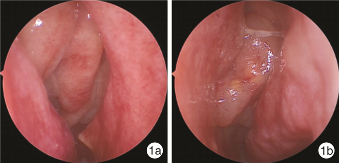
- Figure 1.
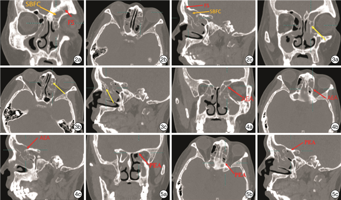
- Figure 2.
- Figure 6.


 下载:
下载:

