Clinical analysis of treatment and postoperative efficacy in neonatal congenital pyriform sinus fistula
-
摘要: 目的 探讨新生儿先天性梨状窝瘘(CPSF)的诊断与治疗方法。方法 回顾性分析5例新生儿CPSF手术治疗的临床资料, 分析患儿的临床症状、辅助检查、手术方式, 术后进行阶段性随访。结果 5例患儿均已明确诊断并顺利完成手术, 术后无咽瘘、吞咽困难、内瘘口周围及瘘管远端感染。所有患儿术后随访3个月~2年, 无一例复发。结论 新生儿CPSF较为罕见, 病程短, 病情进展快, 严重时可危及生命, 需及时处理。Abstract: Objective To discuss the diagnosis and treatment of congenital pyriform sinus fistula(CPSF) in newborn.Methods Clinical data of 5 patients with CPSF innewborn were reviewed and the clinical symptoms, auxiliary examinations, surgical methods were analyzed after the operation, patients were followed up closely at different stages.Results All the 5 neonates successfully completed the surgery without pharyngeal fistula, dysphagia, perifistula and distal fistula infection. Follow-up survey ranged from 3 months to 2 years and no one recurred.Conclusion Neonatal CPSF is a rare disease with a short course of disease and rapid progression. In severe cases, it may threaten life and should be treated in time.
-
Key words:
- neonate /
- pyriform sinus fistula /
- surgical procedures, operative
-

-
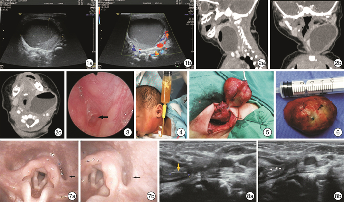
图 1 颈部彩超检查 1a:左甲状腺形态失常,其深方探及一个无回声区,范围5.19 cm×3.7 cm×2.8 cm,边界清,可见气体样强回声斑;1b:内部未探及明显血流信号,周边与甲状腺关系密切,考虑甲状腺囊肿合并内出血? 图 2 颈部CT检查 2a:CT矢状位示左侧颈部低密度肿块,边界清晰,大小3.6 cm×3.1 cm×3.2 cm,内见气液平,薄层囊壁可见强化;病灶上至左侧腮腺下缘、下至锁骨胸骨端上缘,未越过中线;2b:CT冠状位示腮腺、颌下腺、咽腔、左侧梨状窝、甲状腺左叶受压变形右移;2c:CT水平位示左颈部巨大囊性含气占位,与周围结构分界不清; 图 3 支撑喉镜检查 5例患儿最终均在支撑喉镜下显露左侧梨状窝底部内瘘口并确诊,梨状窝周围黏膜明显水肿(箭头所示); 图 4 1例患儿入院后出现呼吸困难,即刻行颈部穿刺抽吸CPSF囊肿,抽出淡黄色液体16 mL; 图 5 术中所见 术中见囊性肿物边界清楚,易与周围组织分离,囊壁较厚,分离至基底部呈瘘管状,见亚甲蓝标记; 图 6 完整切除后的肿物 肿物直径约5 cm,呈椭球形,囊壁较厚,表面光滑; 图 7 CPSF术后3个月电子喉镜复查 左侧梨状窝黏膜无水肿(7a),未见瘘口(7b); 图 8 颈部彩超定期复查 8a:左侧梨状窝区域可见管状结构,长0.7 cm,末端闭锁;8b:内无炎症反应及积液,吞咽时无分泌物进入。
-
[1] 中国妇幼保健学会微创分会儿童耳鼻咽喉学组. 儿童先天性梨状窝瘘诊断与治疗临床实践指南[J]. 临床耳鼻咽喉头颈外科杂志, 2020, 34(12): 1060-1064. https://www.cnki.com.cn/Article/CJFDTOTAL-LCEH202012002.htm
[2] 董锦锦, 田秀芬. 先天性梨状窝瘘的诊断与治疗经验探讨[J]. 中华耳鼻咽喉头颈外科杂志, 2018, 53(6): 444-447. doi: 10.3760/cma.j.issn.1673-0860.2018.06.011
[3] Sheng Q, Lv Z, Xu W, et al. Differences in the diagnosis and management of pyriform sinus fistula between newborns and children[J]. Sci Rep, 2019, 9(1): 18497. doi: 10.1038/s41598-019-55050-9
[4] 陈良嗣, 张思毅, 罗小宁, 等. 先天性第四鳃裂畸形的诊断和治疗[J]. 中华耳鼻咽喉头颈外科杂志, 2010, (10): 835-838. doi: 10.3760/cma.j.issn.1673-0860.2010.10.012
[5] 刘大波, 钟建文, 黄振云. 临床小儿耳鼻喉疾病诊疗学[M]. 北京: 科学技术文献出版社, 2017: 434-435.
[6] 盛晓丽, 陈良嗣, 许咪咪, 等. 儿童双侧先天性梨状窝瘘1例[J]. 临床耳鼻咽喉头颈外科杂志, 2020, 34(9): 848-850. https://www.cnki.com.cn/Article/CJFDTOTAL-LCEH202009018.htm
[7] Chen T, Chen J, Sheng Q, et al. Pyriform Sinus Fistula in the Fetus and Neonate: A Systematic Review of Published Cases[J]. Front Pediatr, 2020, 25(8): 502-502.
[8] Chaudhary N, Gupta A, Motwani G, et al. Fistula of the fourth branchial pouch[J]. Am J Otolaryngol, 2003, 24: 250-252. doi: 10.1016/S0196-0709(03)00026-7
[9] Liu Z, Han J, Fu F, et al. How to make an accurate diagnosis of fetal pyriform sinus fistula in utero: experience at a single medical center in mainland China[J]. Eur J Obstet Gynecol Reprod Biol, 2018, 228: 76-81. doi: 10.1016/j.ejogrb.2018.05.039
[10] Rossi ME, Moreddu E, Leboulanger N, et al. Fourth branchial anomalies: Predictive factors of therapeutic success[J]. J Pediatr Surg, 2019, 54(8): 1702-1707. doi: 10.1016/j.jpedsurg.2019.02.005
[11] 刘菊仙, 邱逦, 文晓蓉, 等. 小儿淋巴管瘤超声及临床病理特点[J]. 四川大学学报(医学版), 2017, 48(6): 949-952, 962. https://www.cnki.com.cn/Article/CJFDTOTAL-HXYK201706033.htm
[12] Nair SC. Vascular Anomalies of the Head and Neck Region[J]. J Maxillofac Oral Surg, 2018, 17(1): 1-12. doi: 10.1007/s12663-017-1063-2
[13] Trappey AF 3rd, Hirose S. Esophageal duplication and congenital esophageal stenosis[J]. Semin Pediatr Surg, 2017, 26(2): 78-86. doi: 10.1053/j.sempedsurg.2017.02.003
[14] Hosokawa T, Yamada Y, Takahashi H, et al. Optimal Timing of the First Barium Swallow Examination for Diagnosis of Pyriform Sinus Fistula[J]. AJR Am J Roentgenol, 2018, 211(5): 1122-1127. doi: 10.2214/AJR.18.19841
[15] Laababsi R, Elbouhmadi K, Bouzbouz A, et al. Misdiagnosed pyriform sinus fistula revealed by iterative neck abscesses: A case report and review of the literature[J]. Ann Med Surg(Lond), 2020, 59: 64-67. doi: 10.1016/j.amsu.2020.08.051
[16] Li Y, Lyu K, Wen Y, et al. Third or fourth branchial pouch sinus lesions: a case series and management algorithm[J]. J Otolaryngol Head Neck Surg, 2019, 48(1): 61-61. doi: 10.1186/s40463-019-0371-6
[17] Pan J, Zou Y, Li L, et al. Clinical and imaging differences between neonates and children with pyriform sinus fistula: which is preferred for diagnosis, computed tomography, or barium esophagography?[J]. J Pediatr Surg, 2017, 52(11): 1878-1881. doi: 10.1016/j.jpedsurg.2017.08.006
[18] Leboulanger N, Ruellan K, Nevoux J, et al. Neonatal vs delayed-onset fourth branchial pouch anomalies: therapeutic implications[J]. Arch Otolaryngol Head Neck Surg, 2010, 136(9): 885-890. doi: 10.1001/archoto.2010.148
[19] Teng Y, Huang S, Chen G, et al. Congenital pyriform sinus fistula presenting as a neck abscess in a newborn: A case report[J]. Medicine(Baltimore), 2019, 98(44): e17784.
[20] Mou JW, Chan KW, Wong YS, et al. Recurrent deep neck abscess and piriform sinus tract: a 15-year review on the diagnosis and management[J]. J Pediatr Surg, 2014, 49(8): 1264-1267. doi: 10.1016/j.jpedsurg.2013.10.018
[21] 郭宇峰, 高兴强, 邓海燕. 婴幼儿先天性梨状窝瘘支撑喉镜下内镜辅助低温等离子射频消融术疗效分析[J]. 临床耳鼻咽喉头颈外科杂志, 2020, 34(5): 455-458. https://www.cnki.com.cn/Article/CJFDTOTAL-LCEH202005018.htm
[22] 鲁秀敏, 桑建中, 张亚民, 等. 低温等离子治疗先天性梨状窝瘘57例[J]. 中国微创外科杂志, 2020, 20(8): 730-733. doi: 10.3969/j.issn.1009-6604.2020.08.014
[23] Arunachalam P, Vaidyanathan V, Sengottan P. Open and Endoscopic Management of Fourth Branchial Pouch Sinus-Our Experience[J]. Int Arch Otorhinolaryngol, 2015, 19(4): 309-313. doi: 10.1055/s-0035-1556823
[24] 纪尧峰, 赵振鹿, 周钦, 等. 低温等离子微创治疗儿童梨状窝瘘[J]. 临床耳鼻咽喉头颈外科杂志, 2019, 33(5): 461-463. https://www.cnki.com.cn/Article/CJFDTOTAL-LCEH201905020.htm
-


 下载:
下载: