
Analysis of correlation between the pneumatization degree of paranasal sinuses and skull base and spontaneous cerebrospinal fluid rhinorrhea
-
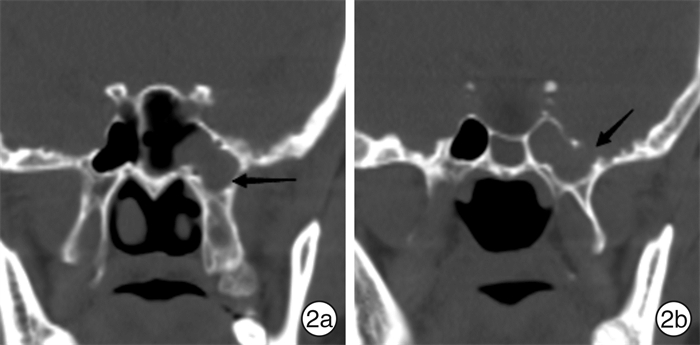
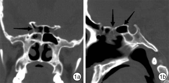
摘要: 目的 探讨鼻窦及颅底气化程度与自发性脑脊液鼻漏(SCSFR)发病之间的关系。方法 回顾性分析107例SCSFR患者的临床资料, 观察并评估CT平扫所表现的鼻窦及颅底气化程度, 比较分析包括额气房、蝶筛气房(Onodi气房)、蝶窦外侧隐窝、前床突气化和后床突气化在内的影像学特征的有无及分型在SCSFR组(病例组)和鼻中隔偏曲组(对照组)的差异。结果 107例SCSFR患者, 共发现瘘口108处, 好发部位为筛窦42处(38.89%), 其次为嗅裂40处(37.04%), 蝶窦23处(21.30%), 额窦3处(2.77%)。病例组Onodi气房发生率高于对照组, 差异有统计学意义(χ2=4.755, P < 0.05), 分级与SCSFR的发生存在线性关系(Z=3.345, P < 0.05), Onodi气房的级别越高, SCSFR发病的可能性越高。对于蝶窦外侧隐窝, Ⅲ型蝶窦外侧隐窝的发生率病例组显著高于对照组(χ2=18.725, P < 0.05), 蝶窦外侧隐窝的分级与SCSFR的发生存在线性关系(Z=3.578, P < 0.05), 蝶窦外侧隐窝气化的级别越高, SCSFR发病的可能性越高。病例组额气房的发生率与对照组比较, 差异无统计学意义(χ2=0.672, P>0.05), 其分级与SCSFR的发病之间不存在线性关系(Z=0.503, P>0.05)。病例组前床突气化及后床突气化与对照组比较, 差异无统计学意义(P>0.05), 二者分级与SCSFR之间均不存在线性关系(P>0.05)。结论 SCSFR的最好发部位为筛窦, 其次为嗅裂; 蝶窦外侧隐窝过度气化和Onodi气房的发生与SCSFR发病密切相关。Abstract: Objective To investigate the relationship between the pneumatization degree of paranasal sinuses and skull base and the incidence of spontaneous cerebrospinal fluid rhinorrhea (SCSFR).Methods Date of 107 patients with SCSFR were reviewed retrospectively. Using CT scans, investigator evaluated the pneumatization degree of paranasal sinuses and skull base, and compared classifications of various imaging characteristics between SCSFR group(case group) and nasal septum deviation group(control group), including frontal cells, Onodi cell, lateral recess of sphenoid sinus (LRSS), pneumatization of anterior clinoid process and pneumatization of posterior clinoid process.Results One hundred and eight fistulas are found among 107 cases. The most common site of the fistulas is found in ethmoid sinus (38.89%), followed by olfactory cleft(37.04%), sphenoid sinus (21.30%) and frontal sinus (2.77%).The incidence of Onodi cell was significantly different between case and control group(χ2=4.755, P < 0.05), and the classification of Onodi cell has a linear effect on the occurrence of SCSFR (Z=3.345, P < 0.05), which suggests the increasing level of Onodi cell can increase the probability in occurrence of SCSFR. The incidence of type Ⅲ LRSS in the case group is significantly higher than in the control group(χ2=18.725, P < 0.05), a linear correlation is found between the classification of LRSS and the incidence of SCSFR (Z=3.578, P < 0.05).There was no significant difference in the incidence of frontal cells between the two groups(χ2=0.672, P>0.05), nor was there a linear relationship between the classification of frontal cells and the incidence of SCSFR (Z=0.503, P>0.05). Pneumatization of anterior clinoid process and posterior clinoid process were no significant difference between case and control (P>0.05), nor were there linear relationships between their classifications and SCSFR (P>0.05).Conclusion The most common site of SCSFR is ethmoid sinus, followed by olfactory cleft, the hyperpneumatization of the LRSS and Onodi cell are closely related to its pathogenesis.
-

-
表 1 发生在筛窦的SCSFR的相关解剖学特征
例(%) 影像学特征 筛窦组 对照组 χ2 P Onodi气房 无 29(69.05) 85(85.00) 4.755 0.029 Ⅰ型 4(9.52) 13(13.00) 0.339 0.560 Ⅱ型 5(11.91) 1(1.00) 6.205 0.013 Ⅲ型 4(9.52) 1(1.00) 4.065 0.044 额气房 无 26(61.91) 69(69.00) 0.672 0.412 Ⅰ型 13(30.95) 25(25.00) 0.535 0.465 Ⅱ型 2(4.76) 2(2.00) 0.124 0.725 Ⅲ型 1(2.38) 4(4.00) 0 1 Ⅳ型 0 0(0) - 1  下载: 导出CSV
表 2 发生在蝶窦的SCSFR的相关解剖学特征
例(%) 影像学特征 蝶窦组 对照组 χ2 P 蝶窦外侧隐窝 Ⅰ型 2(8.70) 18(18.00) 0.604 0.437 Ⅱ型 5(21.74) 59(59.00) 10.402 0.001 Ⅲ型 16(69.57) 23(23.00) 18.725 0 前床突气化 无 21(91.30) 90(90.00) 0 1 Ⅰ型 2(8.70) 2(2.00) - 0.158 Ⅱ型 0(0) 6(6.00) 0.446 0.504 Ⅲ型 0(0) 2(2.00) - 1 后床突气化 无 23(100.00) 100(100.00) - 1 Ⅰ型 0(0) 0(0) - - Ⅱ型 0(0) 0(0) - - Ⅲ型 0(0) 0(0) - -
下载: 导出CSV
-
[1] Mikayilli M, Hasanov T, Otluoglu GD, et al. Spontaneous Idiopathic Cerebrospinal Fluid Rhinorrhea[J]. J Craniofac Surg, 2019, 30(7): 2265-2267. doi: 10.1097/SCS.0000000000005992
[2] Englhard AS, Volgger V, Leunig A, et al. Spontaneous nasal cerebrospinal fluid leaks: management of 24 patients over 11 years[J]. Eur Arch Otorhinolaryngol, 2018, 275(10): 2487-2494. doi: 10.1007/s00405-018-5089-y
[3] Lobo BC, Baumanis MM, Nelson RF. Surgical repair of spontaneous cerebrospinal fluid(CSF)leaks: A systematic review[J]. Laryngoscope Investig Otolaryngol, 2017, 2(5): 215-224. doi: 10.1002/lio2.75
[4] Keshri A, Jain R, Manogaran RS, et al. Management of Spontaneous CSF Rhinorrhea: An Institutional Experience[J]. J Neurol Surg B Skull Base, 2019, 80(5): 493-499. doi: 10.1055/s-0038-1676334
[5] TAME K, GILBERTA L. Spontaneous cerebrospinal fluid leak and idiopathic intracranial hypertension[J]. Curr Opin Ophthalmol, 2019, 30: 467-471. doi: 10.1097/ICU.0000000000000603
[6] 赵薇, 冀永进, 安云芳, 等. 内镜经翼突入路处理蝶窦外侧隐窝病变4例(附文献复习)[J]. 临床耳鼻咽喉头颈外科杂志, 2019, 33(5): 411-415. https://www.cnki.com.cn/Article/CJFDTOTAL-LCEH201905008.htm
[7] Johari HH, Mohamad I, Sachlin IS, et al. A computed tomographic analysis of frontal recess cells in association with the development of frontal sinusitis[J]. Auris Nasus Larynx, 2018, 45(6): 1183-1190. doi: 10.1016/j.anl.2018.04.010
[8] Özdemir A, Bayar Muluk N, Asal N, et al. Is there a relationship between Onodi cell and optic canal?[J]. Eur Arch Otorhinolaryngol, 2019, 276(4): 1057-1064. doi: 10.1007/s00405-019-05284-0
[9] Burulday V, Muluk NB, Akgül MH, et al. Presence and types of anterior clinoid process pneumatization, evaluated by Multidetector Computerized Tomography[J]. Clin Invest Med, 2016, 39(3): E105-110. doi: 10.25011/cim.v39i3.26799
[10] Yazici D. The effect of frontal sinus pneumatization on anatomic variants of paranasal sinuses[J]. Eur Arch Otorhinolaryngol, 2019, 276(4): 1049-1056. doi: 10.1007/s00405-018-5259-y
[11] Burulday V, Akgül MH, Muluk NB, et al. Evaluation of posterior clinoid process pneumatization by multidetector computed tomography[J]. Neurosurg Rev, 2017, 40(3): 403-409. doi: 10.1007/s10143-016-0794-8
[12] Keshri A, Jain R, Manogaran RS, et al. Management of Spontaneous CSF Rhinorrhea: An Institutional Experience[J]. J Neurol Surg B Skull Base, 2019, 80(5): 493-499. doi: 10.1055/s-0038-1676334
[13] Gharzouli I, Verillaud B, Tran H, et al. Value of systematic analysis of the olfactory cleft in case of cerebrospinal rhinorrhea: incidence of olfactory arachnoid dilatation[J]. Eur Arch Otorhinolaryngol, 2016, 273(7): 1643-1647. doi: 10.1007/s00405-015-3599-4
[14] Song X, Wang D, Sun X, et al. Endoscopic Repairs of Sinonasal Cerebrospinal Leaks: Outcome and Prognostic Factors[J]. J Craniofac Surg, 2018, 29(1): 182-187. doi: 10.1097/SCS.0000000000004175
[15] Majhi S, Sharma A. Outcome of Endoscopic Cerebrospinal Fluid Rhinorrhoea Repair: An Institutional Study[J]. Indian J Otolaryngol Head Neck Surg, 2019, 71(1): 76-80. doi: 10.1007/s12070-018-1485-2
[16] 郭艺芳. 我国高血压防控现状的最新数据与启示[J]. 中华高血压杂志, 2016, 24(6): 504. https://www.cnki.com.cn/Article/CJFDTOTAL-ZGGZ201606003.htm
[17] 张璞, 穆俊晌. 颅内压增高与自发性脑脊液鼻漏[J]. 中华耳鼻咽喉头颈外科杂志, 2015, 50(10): 870-872.
[18] Scott KW, Tavanaiepour D, Tavanaiepour K, et al. Spontaneous Cerebral Spinal Fluid Rhinorrhea with Meningocele Secondary to Congenital Foramen Rotundum Defect: A Case Report and Review of Literature[J]. Asian J Neurosurg, 2020, 15(1): 187-189. doi: 10.4103/ajns.AJNS_228_19
[19] Aggarwal V, Nair P, Shivhare P, et al. A Case of Evolving Bilateral Sphenoidal Meningoencephaloceles: Case Report and Review of the Literature[J]. World Neurosurg, 2017, 100: 708, e11-708. e17. doi: 10.1016/j.wneu.2017.02.037
[20] 董继永, 杨本涛, 鲜军舫. 成人蝶窦自发性脑脊液鼻漏及并存颅内高压的影像表现[J]. 中华放射学杂志, 2016, 50(1): 8-12.
[21] Rajasekar G, Nair P, Abraham M, et al. Cerebrospinal fluid rhinorrhea from the lateral recess of sphenoid sinus: More to it than meets the eye[J]. Neurol India, 2019, 67(1): 201-206.
[22] Illing E, Schlosser RJ, Palmer JN, et al. Spontaneous sphenoid lateral recess cerebrospinal fluid leaks arise from intracranial hypertension, not Sternberg's canal[J]. Int Forum Allergy Rhinol, 2014, 4(3): 246-250. doi: 10.1002/alr.21262
[23] Harley B, Wickremesekera A, Tan N, et al. Endoscopic transnasal repair of two cases of spontaneous cerebrospinal fluid fistula in the foramen rotundum[J]. J Clin Neurosci, 2019, 59: 350-352. doi: 10.1016/j.jocn.2018.10.020
-
图(2)
表(2)
计量
- 文章访问数: 1558
- PDF下载数: 477
- 施引文献: 0